


- Protocols

- Articles and Issues
- For Authors
- About
- Become a Reviewer


Articles in press
Articles In Press
Articles In Press are peer reviewed and have been accepted for publication. Please note that these versions may be subject to further edits before their final online publication. Nevertheless, Articles In Press are citable using the DOI. Upon the formal online publication, the article will no longer be listed here, but existing links will automatically redirect to the final version in the corresponding issue.
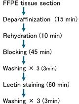
Visualizing Cell Type–Specific Glycan Profiles in Tissue Sections With Glycan Painting
Glycans play critical roles in numerous biological processes. Therefore, visualizing the spatial distribution of glycans within cells and tissues is essential for understanding tissue architecture and cellular identity. Glycan painting is a novel tissue-staining method that employs three fluorescently labeled lectins with different glycan-binding specificities to visualize cell type–specific glycan profiles as unique color patterns. In this study, the protocol was validated using formalin-fixed paraffin-embedded sections of mouse embryos. Although Glycan painting is a qualitative method, each cell population that is difficult to distinguish with a single lectin can be differentiated as unique color patterns generated from the combined images of three fluorescent lectins. Moreover, because it does not require costly reagents or specialized equipment, Glycan painting provides a cost-effective and broadly accessible approach for full-color tissue imaging and cell-type discrimination.
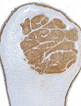
Mouse Intratongue Injection With Cancer Cells—An Orthotopic Model for Head and Neck Squamous Cell Carcinoma
Syngeneic and orthotopic injection of cancer cells in mice is a gold standard model in pre-clinical cancer research. Herein, we detail a protocol of orthotopic intratongue injection as a model for upper respiratory tract cancer studies. This model recapitulates tumor growth and invasion in an orthotopic site, the oral cavity mucosa. It can be experimentally designed in a syngeneic context or used to study patient-derived cells in immunocompromised animals. Procedurally, mice are anesthetized and injected with a small volume of cancer cell suspension near the lateral border of the tongue, right under the mucous membrane. Post-injection, animal welfare is carefully attended to by appropriate analgesia, while providing soft food for the duration of the experiment to ensure the ability of the mice to eat ad libitum. Tongues and potential metastatic organs are dissected and can be sampled and analyzed using relevant state-of-the-art methods, including -omics and imaging approaches. We describe a broad range of distinct tumor profiling and analysis methods applicable to this model. These include physical measurements of elasticity with atomic force microscopy on fresh tissue, hematoxylin-eosin staining and standard immunohistochemistry, and confocal microscopy applied to paraffin-embedded tongue sections. This model is orthotopic, accessible without surgery, easy to monitor, and rapid to perform.
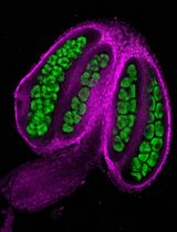
Anther Dissection and Confocal Imaging of Developing and Mature Pollen in Arabidopsis
Pollen development is a fundamental process underlying plant fertility that involves a series of coordinated cellular transitions from microspore mother cells (MMCs) to mature pollen grains. Conventional approaches for studying this process often require tissue sectioning, extensive sample preparation, and specialized instrumentation operated by trained personnel, making them labor-intensive, time-consuming, costly, and poorly suited for high-throughput analysis. Here, we describe simpler protocols for preparing and imaging both developing pollen lineage cells and mature pollen grains from Arabidopsis. By combining targeted anther dissection with gentle extrusion of pollen cells from anthers, fluorescent staining, and confocal microscopy, our protocol makes pollen cells at different developmental stages accessible for high-resolution imaging, enabling visualization of cellular morphology and protein localization, as well as fluorescence-based protein quantification. We also describe a complementary workflow for imaging mature pollen grains that provides a rapid alternative to scanning electron microscopy (SEM) for examining pollen surface structures and patterns. Together, these protocols offer practical approaches for investigating the cellular and molecular processes underlying pollen development and for characterizing the surface morphology of mature pollen.
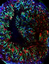
Human iPSC Line SCTi003-A Differentiation Into Kidney Organoids
Kidney organoids derived from human induced pluripotent stem cells (hiPSCs) have emerged as valuable tools for studying human kidney development, disease mechanisms, and therapeutic responses. However, widespread application of these models has been limited by variability in differentiation efficiency, organoid composition, reproducibility, and scalability across existing protocols. Robust and standardised methods are therefore needed to improve experimental consistency and facilitate wider adoption of kidney organoid systems in basic and translational research. Here, we describe an optimised and reproducible protocol for differentiating a commercially available hiPSC SCTi003-A line into kidney organoids (KiO). The protocol builds upon earlier kidney differentiation frameworks and involves directed induction of the posterior primitive streak, intermediate mesoderm specification, and subsequent three-dimensional organoid assembly. Through systematic optimisation of differentiation media, CHIR99021 concentration and timing, growth factor supplementation, and culture format, we established a standardised workflow that yields reproducible nephron and collecting duct structures. The protocol is compatible with air–liquid interface (ALI) culture and scalable suspension-based “mini-organoid” (mKiO) and monolayer culture formats that support high-content and high-throughput applications. This validated protocol provides a reliable and accessible platform for generating human KiO, addressing key limitations of reproducibility, scalability, and assay compatibility in existing approaches, and supporting future studies of kidney development, inherited kidney diseases, and preclinical drug testing.
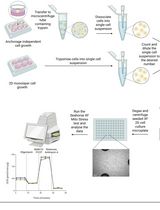
A Dissociation-Based Seahorse XF Workflow for Quantifying Averaged Bioenergetics in Anchorage-Independent Cancer Cell Clusters
Cell clustering during anchorage-independent growth (AIG) is a key hallmark of malignant progression, supporting survival under detachment during metastatic dissemination. However, bioenergetic profiling of cell clusters grown under AIG conditions remains technically challenging, in part because of steep nutrient and oxygen gradients and irregular cell cluster shapes that complicate Seahorse XF assay normalization. Here, we present a dissociation-based Seahorse XF96/XF Pro method that enables high-throughput quantification of the average bioenergetic state of anchorage-independent cancer cell clusters. In this workflow, clusters generated in ultra-low adherent plates are enzymatically and mechanically dissociated into a single-cell suspension immediately prior to Seahorse XF Mito Stress Test analysis on poly-D-lysine-coated 2D microplates. This approach ensures that all viable cells throughout each cluster contribute equally to oxygen consumption rate (OCR) measurements and reduces the need for complex post hoc normalization. We further apply this workflow to an in vitro metastasis model in which A549 lung cancer cells are studied as attached, detached (anchorageindependent), and reattached populations, mimicking primary tumor cells, circulating/clustered cells, and secondary lesions, respectively. Detached cells exhibit reduced mitochondrial membrane potential and diminished basal and maximal OCR, whereas reattached cells largely restore membrane potential and respiratory capacity to levels comparable to attached cells, demonstrating that detachment-induced mitochondrial dysfunction is reversible upon reattachment. These findings align with previous work showing that anchorage-independent and clustered cancer cells undergo reversible metabolic reprogramming, including suppression of oxidative metabolism, engagement of reductive pathways to support redox homeostasis, and altered ROS handling to facilitate metastatic colonization. Collectively, this dissociation-based Seahorse workflow provides a practical and accessible platform to interrogate metabolic adaptations to anchorage-independent growth and cell clustering and identify bioenergetic vulnerabilities of metastasis-relevant cancer cell states.
A Batch-Processing Pipeline for RNA-seq Gene Abundance Estimation
RNA sequencing (RNA-seq) datasets provide valuable opportunities to investigate gene and transcript abundance across species, tissues, developmental stages, and experimental conditions. However, processing multiple datasets consistently remains challenging because sequencing runs may differ in library layout, read length, sequencing depth, metadata quality, and analytical settings. Existing RNA-seq workflows often depend on dedicated workflow managers, substantial computational infrastructure, or advanced bioinformatics expertise, which may limit their accessibility for routine analyses. We present a modular Bash- and Python-based batch-processing pipeline for estimating gene and isoform abundance and generating expression matrices from multiple RNA-seq runs. Using an SRA RunTable, a reference genome, and its corresponding gene annotation, the workflow automates reference preparation, sequencing-data retrieval, quality assessment, read preprocessing, alignment, abundance estimation, and post-processing. It produces gene-level TPM and FPKM matrices, individual gene- and isoform-level RSEM outputs, quality-control summaries, and integrated MultiQC reports. Configurable computational resources, sample-level status tracking, automatic download retries, selective reprocessing of failed samples, and controlled removal of intermediate files allow interrupted analyses to resume without repeating completed runs while reducing storage requirements. By combining batch processing, transparent configuration, and restartable execution in a lightweight workflow, this protocol provides an accessible approach for standardized RNA-seq abundance estimation.
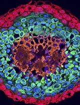
Microtube-Assisted Paraffin Embedding and Sectioning of Fragile 3D Tumor Spheroids for High-Quality Histology


Three-dimensional (3D) spheroid models have become essential in cancer biology, drug screening, and tissue engineering. However, their small size, fragile structure, and tendency to disintegrate during routine histoprocessing present persistent technical challenges. Conventional paraffin embedding often results in tissue fragmentation, loss of spatial orientation, and poor section quality, whereas cryosectioning often compromises cellular morphology. Here, we present a robust, cost-effective protocol for preserving and sectioning fragile 3D spheroids, resulting in high-quality histological sections with intact architecture and excellent cellular detail. The method involves optimized handling and embedding procedures that stabilize spheroids during standard formalin fixation, paraffin infiltration, and microtomy, eliminating mechanical distortion and preserving spherical integrity for consistent sectioning. We demonstrate the successful application of this protocol across different tumor spheroids derived from distinct tissue types, including MDA-MB-231 (breast carcinoma), Caco-2 (colorectal adenocarcinoma), and A549 (lung carcinoma) cell lines, with subsequent compatibility with hematoxylin and eosin (H&E) staining protocols. This protocol is compatible with immunohistochemistry (IHC) and immunofluorescence (IF). In our qualitative comparison with conventional methods, we found that our approach effectively limits sample loss, promotes consistent inter-section reproducibility, and preserves fine structural features, such as necrotic cores, proliferative zones, and extracellular matrix components. This protocol provides a reliable, accessible solution for routine histological analysis of fragile 3D spheroids, facilitating more accurate morphological assessment in translational research settings.
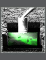
Preparation of Moss Tissue for Cryo-Electron Tomography by High-Pressure Freezing, Focused Ion Beam Milling, and Lift-Out

Cryo-electron tomography (cryo-ET) is a powerful imaging technique that allows visualizing the molecular landscape within cells. In contrast to traditional preparations for electron microscopy deploying chemical fixation and contrasting agents, cryo-ET directly images cryo-fixed, i.e., vitrified samples, providing a faithful and near-native representation of cellular ultrastructure and its macromolecular constituents. While cryo-ET in combination with thinning by focused ion beam (FIB) milling is well established for single small or thin cells that can be vitrified by plunge freezing, its application to tissues is still challenging in terms of vitrification and thinning strategies. Intact plant tissues are particularly difficult, due to their relatively large cell size and the presence of vacuoles, which often occupy a large fraction of cells and usually do not vitrify. Here, we describe two methods for vitrification of the filamentous tissue (protonemata) of moss (Physcomitrium patens) by two high-pressure freezing (HPF) approaches. We also describe in detail how to prepare thin sections (lamellae) suitable for cryo-ET data acquisition by either on-grid FIB milling or lift-out with three different attachment methods that can be applied depending on skill and experimental setup of users. For the preparation of lift-out lamellae, we introduce an additional trimming step that can help to improve lamella quality. We illustrate how subcellular structures can be targeted by aiming for cell junctions using cryo-fluorescence light microscopy. The different preparation procedures can be used for samples of different sizes and other multicellular systems.
Key features
• This protocol builds on serial lift-out [1] but was adapted for moss and can be applied to other species and tissues, as described in [2].
• We describe two high-pressure freezing approaches for different specimen sizes and procedures for sample screening and targeted lamella preparation aided by cryo-fluorescence light microscopy.
• We describe on-grid lamella preparation and three lift-out approaches with a novel, more robust attachment mode.
• By applying an additional trimming step for lift-out sections, this protocol was optimized for lamella quality rather than quantity.
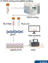
Suspension-Based Intracellular Immunofluorescence Staining Coupled With Genome-Wide CRISPR-Cas9 Screening to Identify Regulators of O-GlcNAcylation
Protein O-GlcNAcylation is a dynamic and reversible post-translational modification that regulates diverse cellular processes, including transcription, signal transduction, metabolism, and cell fate determination. Systematic identification of genes that modulate global O-GlcNAc levels remains technically challenging at the genome scale. Here, we describe a pooled CRISPR-Cas9 screening protocol that combines the human genome-scale CRISPR knockout (GeCKO) v2 knockout library with intracellular immunofluorescence staining using the anti-O-GlcNAc antibody RL2 and fluorescence-activated cell sorting (FACS). In this workflow, HEK293T cells are transduced with the GeCKO v2 lentiviral library at a low multiplicity of infection to ensure predominantly single-sgRNA integration. Following puromycin selection, cells are fixed, permeabilized, and stained in suspension with RL2. The top 5% of cells with the highest intracellular RL2 fluorescence are collected as the RL2-high population, while a corresponding unsorted/input sample is retained as the reference for downstream sgRNA enrichment analysis. Genomic DNA is recovered from the RL2-high and unsorted/input samples, and integrated sgRNA cassettes are amplified through a two-step PCR and library-preparation workflow for Illumina sequencing. This protocol enables the identification of candidate genes whose knockout is associated with increased intracellular RL2 fluorescence.
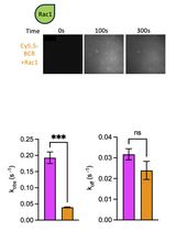
Preparation of Phosphoinositide-Containing Supported Lipid Bilayers and Quantitative Assessment of Protein Recruitment to Membrane Surfaces
Small GTPases and many other peripheral membrane proteins regulate essential cellular processes through dynamic interactions with cellular membranes. These interactions are often controlled by membrane composition, particularly phosphoinositides (PIPs), which modulate the recruitment of GTPases, their regulators, and their effectors. Quantitative characterization of protein recruitment to defined membrane environments remains technically challenging. Here, we describe a protocol for preparing supported lipid bilayers (SLBs) containing defined phosphoinositide compositions in polydimethylsiloxane (PDMS) chambers and monitoring protein recruitment by total internal reflection fluorescence (TIRF) microscopy. Purified fluorescently labeled proteins are incubated with SLBs to measure membrane association in real time under controlled biochemical conditions. The method enables quantitative analysis of membrane recruitment kinetics and comparison of protein binding across different lipid compositions or in the presence of partner proteins. Combining the biochemical precision of a reconstituted membrane system with the sensitivity of TIRF imaging, this protocol provides a robust and versatile platform for studying lipid-dependent membrane recruitment. Although developed to investigate small GTPase signaling, it is readily applicable to a wide range of peripheral membrane proteins and membrane-associated signaling mechanisms.
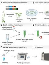
An Optimized Protocol for TurboID-Based Proximity Labeling and Sample Preparation for Mass Spectrometry in Arabidopsis thaliana

Plant proximity labeling proteomics enables the identification of transient and weak intracellular protein interactions that are undetectable via traditional biochemical assays. Conventional enrichment pipelines suffer severe quantitative bias caused by urea-derived chemical artifacts and high mass spectrometry background signals from sample- and carrier-derived impurities. This protocol describes a complete standardized workflow for biotinylated protein extraction, enrichment, and LC-MS/MS sample preparation optimized for Arabidopsis seedlings. The procedure integrates controlled urea dilution and pre-desalting to suppress non-enzymatic protein modifications, introduces acetylation blocking of streptavidin magnetic beads to mitigate streptavidin degradation during on-bead digestion, and applies two-step on-bead trypsin digestion to improve peptide coverage. Multi-point sample retention and dual protein quantification are implemented throughout all experimental stages to ensure full-process quality control. Subsequent data processing pipelines using Spectronaut for data-independent acquisition (DIA) data and MaxQuant for data-dependent acquisition (DDA) data are also detailed for consistent proteome quantification. This workflow delivers higher protein recovery and better signal-to-noise ratios than standard protocols while offering flexible adaptation for various crop proximity labeling and affinity enrichment proteomic research.
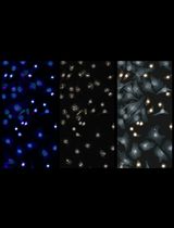
A Python-Based Workflow for Image-Based Single-Cell Phenotypic Profiling From Fluorescence Microscopy Images
While advances in omics technologies have greatly improved our understanding of cellular heterogeneity, there is an increasing need for complementary approaches that capture the spatial organization and structural dynamics of cells. Image-based single-cell phenotypic profiling provides quantitative information on cell morphology and organelle organization, offering valuable insights into cellular function and regulation. Although numerous image analysis tools are available, establishing a complete analysis workflow, from image preprocessing and segmentation to feature extraction and multivariate analysis, often requires substantial computational expertise and software integration. Here, we describe a Python-based workflow for image-based single-cell phenotypic profiling from immunofluorescence microscopy images and provide a detailed protocol for its implementation. Using synchronized HeLa cells with drug-induced mitotic spindle defects as an example, the workflow covers image loading, cell segmentation, quantitative feature extraction, profile integration, dimensionality reduction, clustering, and data visualization. The protocol is accompanied by example datasets, annotated Jupyter Notebooks, and instructions for execution in either a local Python environment or Google Colab, facilitating straightforward implementation and customization. By integrating the entire analysis pipeline within a single coding environment, this workflow enables reproducible and accessible single-cell morphological profiling without requiring specialized imaging equipment or extensive programming expertise. The workflow therefore provides a practical platform for studying cell morphology, organelle organization, and cellular dynamics across a broad range of biological applications.
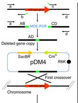
An Improved Method for Rapid Unmarked Gene Deletion in Bacteria: Pseudomonas aeruginosa as an Example
The release and updating of whole-genome sequences of several representatives of the human opportunistic pathogen Pseudomonas aeruginosa have laid the ground for investigating the mechanisms of antibiotic resistance, biofilm formation, and virulence, while also offering opportunities for researchers to find new therapeutic targets to control this bacterium using a functional genomics approach. However, there is still a lack of detailed protocols describing gene inactivation methods in P. aeruginosa, resulting in failures and extra time spent designing in-house protocols. Here, we introduce a rapid, efficient, and unmarked deletion mutagenesis method combining overlap extension PCR, efficient conjugation, and the traditionally used sacB-based counter-selection procedure. Efficient generation of deletion mutants using this detailed protocol can be easily completed in one week using standard lab reagents. Importantly, this method may be adaptable to other bacteria where the sacB-based counter-selection system works.
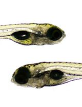
Antiangiogenic Drug Testing Using Proangiogenic and Hypoxia Zebrafish Model
Zebrafish is an excellent in vivo model for high-throughput antiangiogenic drug testing, commonly known as the zebrafish angiogenesis assay. Conventional zebrafish angiogenesis assays are performed in wild-type zebrafish to evaluate the vascular changes in intersegmental and subintestinal vessel regions of the zebrafish larvae at 2 and 3 dpf stages, respectively. However, wild-type zebrafish larvae do not adequately mimic the hypoxia microenvironment and ectopic vessel branching characteristics of cancer. To overcome this limitation, we developed a genetically engineered zebrafish model with constitutive activation of the hypoxia signaling pathway by targeting the vhl, a tumor suppressor, by negative regulation of the hypoxia pathway, using CRISPR mutagenesis. This model exhibits robust ectopic blood vessel branching throughout the larval body, thereby recapitulating pathological angiogenesis. The utility of this zebrafish model system for drug screening was validated with the sorafenib treatment, a known antiangiogenic tyrosine kinase inhibitor. Overall, this proangiogenic hypoxia zebrafish model provides a physiologically relevant platform for testing antiangiogenic drugs.
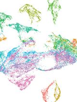
Serial Cryosectioning for the Spatial Transcriptomics of Plant Tissues
Spatial transcriptomics enables genome-wide gene expression profiling while preserving tissue architecture, making it a powerful approach for studying plant developmental transitions. However, preparing small and structurally complex plant tissues for spatial transcriptomics remains technically challenging because samples must be rapidly preserved, precisely oriented, serially sectioned, and accurately positioned within the limited capture area of the Visium slides. Here, we describe an optimized workflow for cryo-embedding, serial cryosectioning, section placement, and data analysis of small plant samples for 10x Genomics Visium spatial transcriptomics. Using maize seedling shoot apices as the target, this protocol includes preparation of custom molds for optimal cutting temperature embedding, rapid fresh sample embedding, serial cryosectioning, section-position marking for Visium HD workflows, and morphological quality assessment of replicate tissue slides before transcript capture. The associated data analysis workflow includes Space Ranger processing, Seurat-based normalization and Harmony integration, anatomical domain annotation, pseudobulk and developmental trend analyses, RNA velocity, pseudotime analysis, transcription factor network analysis, single-cell reference mapping, and 3D transcriptome reconstruction. This computational workflow was developed and tested using maize Visium V1 data, but not Visium HD data. This protocol was used to generate serial spatial transcriptomes of maize shoot apices and developing leaf primordia, enabling reconstruction of gene expression transitions from the shoot apical meristem to sequential leaf developmental stages. The approach is also applicable to other small plant tissues, including Arabidopsis first true leaves and Marchantia thalli.
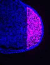
PIC-RNA-seq for Region-Specific Transcriptomic Analysis of Chicken Limb Bud Progenitors
Region-specific RNA sequencing is a powerful approach for investigating tissue differentiation and dynamic changes in gene expression during embryonic development. The chicken embryo has long served as an important model system in developmental biology. However, the limited availability of tissue-specific reporter lines makes region-specific RNA-seq approaches particularly valuable in this organism. Here, we applied photo-isolation chemistry-based RNA sequencing (PIC-RNA-seq) to the somatic lateral plate mesoderm (sLPM) before the emergence of limb bud progenitor cells (LPCs) and to early LPCs in chicken embryos. These analyses revealed the upregulation of multiple genes, including Hox genes, in LPCs, suggesting the initiation of their regional patterning program. This workflow enables visualization of dynamic changes in the gene expression profile of LPCs and should also be applicable to other tissues in avian embryos.
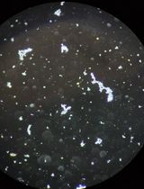
PCR-Guided Isolation of Leptospira Strains From Refrigerated Serum Samples for Serogroup and Genomic Characterization
Leptospirosis is a widespread zoonotic disease caused by pathogenic bacteria of the genus Leptospira. The isolation and comprehensive characterization of circulating strains within a region are essential for understanding the local epidemiology and improving public health surveillance. Historically, whole blood has been the specimen of choice for isolation; however, its efficiency can be limited by the presence of inhibitory substances in the sample, and Leptospira viability may depend on rapid processing and inoculation. Here, we present an in-house culture protocol for the isolation of Leptospira from serum samples previously maintained under refrigeration (i.e., 4–8 °C) for up to 10 days. The protocol employs a real-time PCR-guided strategy by first screening specimens for the lipL32 gene. Positive samples are then inoculated into specialized EMJH media supplemented with AFAS and EMJH+AFAS supplemented with STAFF antibiotic cocktail, followed by incubation at 30 °C for up to six months. Growth is monitored weekly through visual inspection and, once turbid, the presence of Leptospira is determined via dark-field microscopy prior to downstream serogroup and genomic characterization. A significant advantage of this method is the successful recovery of viable Leptospira from non-fresh serum specimens stored under refrigeration, even in samples with low bacterial loads. Additionally, the protocol facilitates broader surveillance by repurposing serum samples already collected for routine serology, increasing the probability of identifying diverse strains without further clinical collection
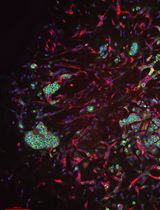
Disease Modeling in iPSC-Derived Human Bone Marrow Organoids
Human bone marrow organoids provide a tractable three-dimensional platform for modeling hematopoiesis and hematologic disease in a human niche–like context. Here, we describe a stepwise protocol for utilizing human induced pluripotent stem cell (iPSC)-derived bone marrow organoids that support autonomous hematopoiesis for hematopoietic disease modeling, mouse xenograft hematopoiesis, and drug sensitivity testing. The workflow combines embryoid body formation, early mesoderm/angiogenic induction under hypoxia, hemogenic endothelial commitment, maturation within a collagen-containing hydrogel, and subsequent suspension culture as individual organoids. The resulting organoids contain endothelial, stromal, and hematopoietic components and reproduce key structural and cellular features of human marrow. We further describe procedures for engraftment of normal donor- or patient-derived CD34+ cells and implantation of mature organoids under the renal capsule of immunodeficient mice to assess in vivo hematopoietic maintenance. In prior applications of this platform, donor-derived CD34+ cells were shown to engraft within the organoid niche and undergo multilineage differentiation, enabling detection of selective erythroid defects caused by DDX41 deficiency and assessment of therapeutic suppression of JAK2V617F-mutant patient-derived hematopoietic cells in a human marrow–like microenvironment. This protocol, therefore, enables disease modeling, in vivo xenograft assessment, and ex vivo functional analysis of patient-derived hematopoietic cells using relatively small input samples.
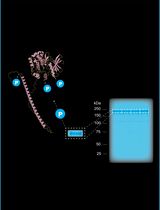
In-Gel Tryptic Digestion and HCD/EThcD LC-MS/MS for Mapping Autophosphorylation Sites
This protocol describes a targeted workflow for identifying and quantitatively comparing kinase autophosphorylation sites, using in-gel proteolytic digestion, LC-MS/MS analysis, phosphosite localisation, and extracted ion chromatogram (XIC) analysis. The detailed protocol begins with an excised SDS-PAGE gel band containing the immunoprecipitated kinase of interest. Standard cell culture, expression, immunoprecipitation, and SDS-PAGE procedures can be used to generate this starting material according to the experimental system under investigation; the specific conditions used for NEK1 are described in our associated research article. Here, the protocol is applied to characterise phosphorylation sites on NEK1—a serine/threonine kinase implicated in amyotrophic lateral sclerosis (ALS)—using a homozygous NEK1 knockout cell background in which GFP-tagged wild-type or kinase-dead NEK1 constructs are re-expressed. This experimental system enables accurate assessment of kinase-dependent phosphorylation events in the absence of endogenous NEK1. A key methodological feature of the workflow is the use of two parallel MS2 fragmentation strategies: higher energy collisional dissociation (HCD) and electron-transfer/HCD (EThcD). EThcD generates complementary b/y and c/z ion series, providing superior localisation confidence for labile phosphorylation events that are difficult to resolve using HCD alone. Relative phosphopeptide abundance across conditions can be assessed using extracted ion chromatograms generated in FreeStyle, or using Skyline software with normalisation to the total ion current (TIC), as performed in the associated NEK1 study. Although demonstrated here using NEK1, the protocol is readily adaptable to other kinases amenable to immunoprecipitation and will be of interest to researchers studying kinase signalling, post-translational modification biology, and disease-associated phosphorylation events.
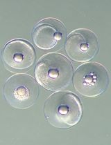
Optimized Phenol–Chloroform–Isoamyl DNA Extraction Protocol for Single Fish Eggs
Reliable DNA extraction is essential for genetic research on marine species; however, obtaining sufficient DNA from single fish eggs remains challenging. Existing protocols often require optimization to achieve high PCR efficiency. The optimized phenol–chloroform–isoamyl extraction protocol presented in this paper improves DNA yield and quality from individual eggs of Atlantic bluefin tuna (Thunnus thynnus), bogue (Boops bops), saddled seabream (Oblada melanura), and painted comber (Serranus scriba) by modifying buffer volumes, incubation times, and washing steps, following prior micropuncturing of eggs on a glass slide. DNA quality is confirmed by spectrophotometry, PCR amplification of the mitochondrial COI gene, electrophoresis, and Sanger sequencing. This method provides a low-cost and effective approach for species identification from individual fish eggs.

Quantitative Colocalization Analysis in Fluorescence Microscopy
Spatial organization of macromolecules is fundamental to cellular function, with colocalization providing key insights into molecular interactions and biological processes. However, quantification remains challenging due to diverse localization patterns and irregular sample geometries. Here, we present a protocol for analyzing colocalization between two fluorescent probes using coAnalyzer, a MATLAB-based software package. coAnalyzer features a user-friendly graphical interface and supports region of interest (ROI) selection, image merging, line scanning, signal isolation, scatterplot generation, and quantitative analysis. coAnalyzer enables colocalization analysis of any two fluorophores, regardless of the proteins or dyes involved. Its broad applicability across a wide range of organisms and sample types demonstrates the robustness, flexibility, and versatility of the platform.
Stereotaxic Injection of Lysophosphatidylcholine Into Mouse Corpus Callosum for Establishment of a Focal Demyelination Model
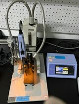
Multiple sclerosis (MS) is a chronic autoimmune disease characterized primarily by inflammatory demyelination of the central nervous system and is one of the leading causes of non-traumatic neurological disability in young and middle-aged adults worldwide. Myelin loss leads to impaired neural conduction, while progressive axonal degeneration resulting from failed remyelination constitutes a major pathological basis for irreversible disability in patients. Among currently approved treatments for MS, effective therapies that directly promote remyelination are still lacking; therefore, establishing animal models that can precisely recapitulate the myelin injury-repair process is essential for elucidating the mechanisms of remyelination and screening remyelination-promoting drugs. Focal demyelination models are important tools for investigating the mechanisms of remyelination and for developing therapeutic strategies for demyelinating diseases such as multiple sclerosis. Unlike the inflammation-driven injury of the experimental autoimmune encephalomyelitis (EAE) model and the systemic metabolic toxicity-induced demyelination of the cuprizone model, the lysophosphatidylcholine (LPC) injection model directly disrupts myelin in the corpus callosum through local injection of a membrane-solubilizing lipid, inducing focal demyelinating lesions and enabling investigators to study, in a controlled manner, the recruitment and differentiation of oligodendrocyte progenitor cells as well as the dynamic process of remyelination. This protocol describes the complete workflow for establishing focal demyelinating lesions by stereotaxic injection of LPC into the mouse corpus callosum, covering surgical preparation, coordinate localization, controlled injection, and postoperative care. Compared with existing methods, its main advantages lie in the precise control of the lesion and the synchronization of the post-injury repair phase, making it highly suitable for quantitative comparisons. Beyond the corpus callosum, this method is also broadly applicable to focal demyelination studies in other white matter tracts (including the spinal cord, optic nerve, and others), serving as a versatile platform for investigating region-specific myelin injury and repair.
Semi-Automated Multiplex Workflow for Functional In Vitro Testing of Chemotherapeutic Treatments in Primary, Patient-Derived Cancer Organoids
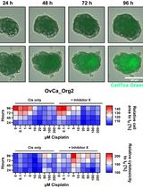
Most existing preclinical models have been limited in their predictive value to mimic patients’ responses, which is a major drawback in drug development and the identification of predictive biomarkers. To overcome these limitations, patient-derived three-dimensional in vitro models have been proposed. One of them is the organoid model, which preserves the original cellular heterogeneity and recapitulates epithelial architecture and functionality. Recently, studies using patient-derived organoids for drug screening applications have increased in quantity, and organoids have already been applied to pancreatic, colon, and lung cancers and female gynecological malignancies. Here, we established a multiplex workflow to analyze longitudinal therapeutic effects of anti-cancer therapeutics on organoid growth, viability, and cytotoxicity by combining state-of-the-art viability measurement with automated live cell imaging. This workflow can be used for the prediction of patient-specific treatment response, high-throughput screening of potential anticancer drugs, and downstream analysis to identify novel therapeutic targets.

Protocol Collections
Comprehensive collections of detailed, peer-reviewed protocols focusing on specific topics



