- Protocols

- Articles and Issues
- For Authors
- About
- Become a Reviewer

Current Issue in 2026
Volume: 16, Issue: 14
Biochemistry
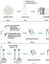
A Practical Experimental Protocol for Identification and Validation of UFMylation Substrate in Human Cells
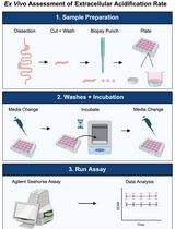
Ex Vivo Assessment of Extracellular Acidification Rate in Murine Intestinal Tissue
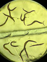
Iodine Staining of Glycogen Storage in Caenorhabditis elegans
Bioinformatics and Computational Biology
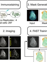
Actin Quantification Using the Filamentous Actin Segmentation Tool (FAST)
Biological Engineering
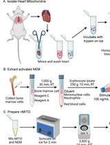
Preparation and Characterization of Neutrophil Membrane-Fused Mitochondria (nMITO)
Cancer Biology
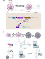
Light-Regulated Cancer Immunotherapy Using Individually Encapsulated Synthetic Circuit–Engineered Cells
Cell Biology
A Universal Resazurin-Based Viability Assay for Prokaryotic and Eukaryotic Cells in 2D and 3D Cultures


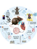
A Simplified Langendorff-Based Method for Mouse Cardiac Myocyte Isolation
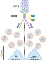
Isolation of Mononucleated and Binucleated Hepatocytes by Flow Cytometry
Developmental Biology
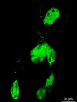
In Vivo Light-Sheet Imaging of Senescence Reporter Activity in a Transparent Killifish
Immunology
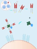
A Streamlined and Time-Saving Approach to Generate HLA-DR15 MHC Class II Tetramers via In Vivo Biotinylation
Microbiology
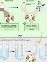
Separating Chromera velia Zoospores From Culture and Estimating Their Average Motility Speed and Lifespan


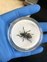
Optimized Field Collection and Gut Dissection Workflows for Microbiome Studies of the Citrus Root Weevil, Diaprepes abbreviatus
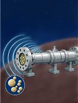
Assessment of Saccharomyces cerevisiae Survival Upon Exposure to Transient High Pressure and Temperature in a High-Intensity Shock Tube for Astrobiology (HISTA)
Molecular Biology
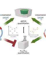
An Accurate and Precise ddPCR-Based Method for Determining the Concentration of Plasmid DNA
Neuroscience
Measuring PINK1 Activity in Single Cells Using a PINK1 Kinase Activity Reporter
Plant Science
Simple Electroporation of Chlamydomonas reinhardtii Strains With an Intact Cell Wall

Gene Editing in Chlamydomonas Using the SCREAM Technique
Stem Cell
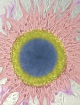
Histological Processing of Organoids for Immunostaining
Satellite Cell Isolation, Culture, and Infection After Retroviral Preparation
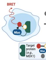
Protocol for Measuring Drug–Target Engagement in Mouse Colorectal Cancer Organoids Using NanoBRET Assay