


- Protocols

- Articles and Issues
- For Authors
- About
- Become a Reviewer


A Modified Slide-Embedded Scanning Electron Microscopy Preparation Method to Visualize Antagonistic Interactions Between Trichoderma viride and Fusarium sp.

(§Technical contact: sandra.tomichen2023@vitstudent.ac.in) Published: Vol 16, Iss 17, Sep 5, 2026 DOI: 10.21769/BioProtoc.5805 Views: 254
Reviewed by: Jessica DavisVandana MishraNidia Maldonado Carmona
 PDF
PDF  Ask a question

Protocol Collections
Comprehensive collections of detailed, peer-reviewed protocols focusing on specific topics




Related protocols
Abstract
Mycoparasitism is an important mechanism of fungal antagonism in which one fungus parasitizes another. This type of interaction plays a major role in the biocontrol activity of Trichoderma spp. against phytopathogenic fungi. Detailed visualization of these interactions is essential for understanding the structural mechanisms involved in fungal antagonism, including hyphal attachment, coiling, penetration, and cellular distortion. Scanning electron microscopy (SEM) is widely used for structural examination of fungal interactions; however, conventional preparation methods such as filter paper systems, membrane overlays, and agar block techniques often result in structural distortion, fragile sample handling, and difficulty in locating defined interaction zones. Here, we describe a modified slide-embedded technique for SEM visualization of mycoparasitic interactions between filamentous fungi. The protocol is adapted from previously reported slide culture approaches and involves embedding pre-cut sterile glass slide fragments directly into potato dextrose agar (PDA), followed by sequential inoculation of Fusarium sp. and Trichoderma viride. Fungal interactions occurring directly on the glass surface are subsequently subjected to fixation with 2.5% glutaraldehyde, graded ethanol dehydration, sputter coating, and SEM observation. Compared with conventional methods, the present approach provides improved handling stability, better preservation of native hyphal architecture, reduced deformation during processing, and easier localization of interaction zones during microscopy. The protocol also enables clear visualization of early antagonistic events such as hyphal coiling, penetration, and surface colonization. Due to its simplicity, reproducibility, and minimal technical complexity, this method serves as a practical and efficient approach for SEM-based investigation of fungal–fungal interactions and can be readily adapted for studying diverse mycoparasitic systems.
Key features
• A simple, cost-effective method to visualize filamentous fungus–fungus interactions, particularly mycoparasitism, using minimal reagents and basic laboratory equipment.
• A standardized inoculation strategy with pathogen inoculation 48 h before the mycoparasite ensures reproducible fungal interaction and mycoparasitic development.
• Blunt-cut square glass slide pieces embedded in PDA enable easy sample handling, storage, transport, and direct preparation for scanning electron microscopy.
• SEM-compatible fixation and ethanol dehydration preserve fungal ultrastructure, enabling high-resolution visualization of hyphal coiling, distortion, and other mycoparasitic interactions.
Keywords: Trichoderma viride Fusarium sp.
Mycoparasitism
Antagonism
Scanning Electron Microscopy
Fusarium sp.
Mycoparasitism
Antagonism
Scanning Electron Microscopy
Graphical overview
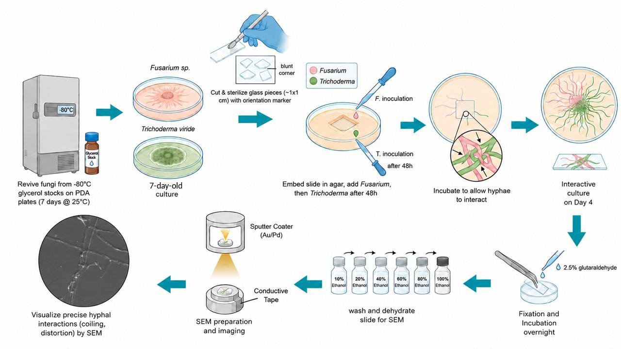
Graphical overview of the slide-embedded technique for scanning electron microscopy (SEM) visualization of mycoparasitic interactions between Trichoderma viride and Fusarium sp. Fungal cultures are revived from glycerol stocks, and a sterile square glass slide piece with a single blunt-cut edge is embedded in potato dextrose agar (PDA). Fusarium sp. is inoculated first and allowed to grow for 48 h, followed by inoculation of T. viride at the advancing hyphal margin. After interaction, the embedded slide is fixed, dehydrated through a graded ethanol series, sputter-coated with Au/Pd, and examined by SEM to visualize mycoparasitic structures such as hyphal coiling and distortion.
Background
Mycoparasitism is one of the major mechanisms underlying fungus–fungus antagonism, in which one living fungus derives nutrients from another living fungal host. Species of Trichoderma are among the most extensively studied mycoparasitic fungi because of their broad host range and strong antagonistic activity against several phytopathogenic fungi [1]. Fungicolous fungi are commonly categorized as symbionts, saprophytes, neutral associates, or mycoparasites depending on the nature of their interaction with other fungi [2,3]. Mycoparasitism is considered an ancestral trait of Trichoderma spp., contributing significantly to their effectiveness as biological control agents against plant pathogens.
In addition to mycoparasitism, Trichoderma spp. exhibit multiple biocontrol mechanisms, including antibiosis, induced systemic resistance (ISR), and competitive exclusion. These fungi behave predominantly as necrotrophs toward their hosts through the secretion of cell wall–degrading enzymes and secondary metabolites that suppress pathogenic fungal growth. Important enzymes produced by Trichoderma spp. include β-glucanases, proteases, cellulases, hemicellulases, and small secreted proteins, whereas secondary metabolites such as gliotoxin contribute to antagonistic activity [4,5].
Visualization of filamentous fungal interactions using scanning electron microscopy (SEM) plays an important role in understanding the structural and mechanistic aspects of mycoparasitism, including hyphal coiling, penetration, attachment, and distortion of host hyphae. Conventionally, SEM studies of fungal interactions have employed filter paper–based systems in which fungi grow over removable substrates prior to fixation and imaging [6]. However, these methods often present several limitations, including fragility of the substrate, difficulty during handling, and distortion of delicate hyphal structures during fixation and dehydration procedures.
To overcome these challenges, alternative techniques such as slide culture methods, cellophane membrane overlays, and coverslip-based systems have been developed to improve structural preservation and facilitate clearer visualization of fungal interaction zones [7,8]. Among these, the slide culture technique is considered one of the most reliable and feasible approaches for studying localized fungal interactions because of its simple setup and improved preservation of fungal morphology [8].
The present protocol describes a modified slide-embedded technique adapted from the novel slide culture approach reported by Bhat 2017 for studying mycoparasitic interactions between filamentous fungi [9]. In this method, pre-cut sterile glass slide fragments are embedded directly into PDA medium, allowing fungal growth and interaction to occur on a stable glass surface suitable for SEM analysis. Compared with conventional methods, this approach offers several advantages: (i) simplified experimental setup without the need for elaborate slide culture assemblies; (ii) improved sample handling and reduced structural deformation during SEM preparation; (iii) enhanced preservation of hyphal interactions; (iv) better visualization of early antagonistic events such as hyphal coiling and penetration; and (v) consistent localization of interaction zones using orientation-marked slide fragments. Consequently, this modified slide-embedded method provides a practical, reproducible, and user-friendly approach for SEM-based visualization of fungal mycoparasitic interactions. In this study, we investigate and visualize the mycoparasitic interaction between Fusarium sp. and the biocontrol fungus Trichoderma viride using SEM.
Materials and reagents
Biological materials
1. Trichoderma viride TVI (Elamala Biotech Lab, Chellarcovil, Kerala)
2. Fusarium sp. isolate STSP (Elamala Biotech Lab, Chellarcovil, Kerala)
Reagents
1. Potato dextrose agar (PDA) (HiMedia, catalog number: MH096)
2. Acetone 99% (Hyma, catalog number: ASA2019)
3. Absolute ethanol 99.9% (MSB Chemical Limited, catalog number: 5268-39); ethanol solutions (10%, 20%, 40%, 60%, and 80%) were prepared in Milli-Q water and syringe-filtered (0.22 μm)
3. Glutaraldehyde 25% aq. solution (SRL, catalog number: 92577)
4. Sodium phosphate dibasic dihydrate (SRL, catalog number: 87258)
6. Sodium phosphate monobasic dihydrate (SRL, catalog number: 40597)
7. Milli-Q water
Solutions
1. 0.1 M phosphate buffer (pH 7.4) (see Recipes)
2. Glutaraldehyde fixative (see Recipes)
Recipes
1. 0.1 M phosphate buffer (pH 7.4)
| Reagent | Final concentration | Quantity or volume |
|---|---|---|
| Sodium phosphate dibasic dihydrate | 75.407 mM | 1.342 g |
| Sodium phosphate monobasic dihydrate | 24.593 mM | 0.383 g |
| Milli-Q water | n/a | Make up to 100 mL |
| Total | n/a | 100 mL |
Dissolve both components in 80 mL of Milli-Q water. Adjust the pH to 7.4 using 1 N HCl or 1 N NaOH and make up the final volume to 100 mL with Milli-Q water. Store at room temperature and chill the required volume to 4 °C before use. This solution can be stored at room temperature for approximately 1–2 weeks if prepared and handled aseptically. Without sterilization (e.g., filtration or autoclaving), it should be used within 24–48 h. When properly sterilized and stored in tightly sealed containers, it remains stable for up to 2 months. Because phosphate buffers are prone to microbial contamination, discard the buffer if cloudiness, particulates, or visible microbial growth is observed. For maximum stability and extended shelf life, store the buffer at 4 °C.
Note: Avoid using KOH to adjust the pH, as excess potassium may alter the buffer composition, disturb the Na+/K+ balance, and affect cellular integrity.
2. Glutaraldehyde fixative
| Reagent | Final concentration | Quantity or volume |
|---|---|---|
| Glutaraldehyde 25% aq. solution | 2.5% | 1 mL |
| 0.1 M phosphate buffer (pH 7.4) | 10 mM | 1 mL |
| Milli-Q water | n/a | 8 mL |
| Total | n/a | 10 mL |
Freshly prepare this solution and then syringe-filter it through a 0.22 μm filter before use. Chill all components to 4 °C prior to preparation (see Troubleshooting 1).
Caution: Toxic if inhaled, ingested, or in contact with skin.
Laboratory supplies
1. Petri dish 100 mm (Borosil Scientific, catalog number: 3160077)
2. Conical flask 500 mL (Borosil Scientific, catalog number: 4980024)
3. Kimtech Science® KimwipesTM (Kimberly-Clark Professional, catalog number: 34155)
4. Inoculation loop (HiMedia, catalog number: LA019)
5. Syringe filter 0.22 μm (Sartorius, catalog number: 16532)
6. Disposable Pasteur pipettes (Genaxy, catalog number: GEN-153)
7. Parafilm M (Amcor, catalog number: PM992)
8. Microtips, 200–1,000 μL (Tarsons, catalog number: 521020)
9. Sterile tissue culture dish 60 mm (Tarsons, catalog number: 960020)
10. Sterile single-use syringe 10 mL
11. Forceps
12. Microscopic glass slide
13. Double-sided adhesive tape
14. Permanent marker
15. Rubber band
16. Newspaper
17. Non-absorbent cotton
Equipment
1. Scanning electron microscope (Carl Zeiss, model: EVO 18 Research)
2. Glass cutter (generic)
3. pH meter (HANNA instruments, model: HI98107)
4. Autoclave (EPS, model: EPS/VA-22)
5. Test tube rack (generic)
6. Laminar air flow (Clean Air Products, model: CAP412)
7. Microbiological incubator (Orbitek, model: LE)
8. Brightfield microscope (Carl Zeiss, model: Axioscope 5)
9. Bunsen burner (generic)
10. Refrigerator (2–8 °C) (generic)
11. Micropipettes 100–1,000 μL (Eppendorf, model: Research Plus Manual Single-channel pipette)
Procedure
A. Revival of fungal cultures from glycerol stocks
1. Prepare PDA according to the manufacturer’s instructions by dissolving the required quantity of medium in distilled water. Sterilize the PDA medium by autoclaving at 121 °C for 15 min.
2. Allow the PDA medium to cool to approximately 40–60 °C.
3. Aseptically pour the molten medium into sterile Petri dishes inside a laminar airflow chamber and allow it to solidify.
4. Inoculate on separate PDA plates with a loopful of conidial suspension (4 × 10⁸ spores mL-1) of Fusarium sp. and Trichoderma viride preserved in 50% glycerol stocks at -80 °C.
5. Incubate the inoculated plates at 25 °C for 7 days to obtain actively growing cultures (Figure 1).
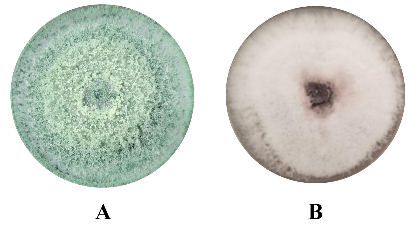
Figure 1. Fully grown fungal culture plates. (A) Seven-day-old culture dish of Trichoderma viride. (B) Seven-day-old culture plate of Fusarium sp.
B. Preparation of glass slide pieces
1. Sterilize a grease-free microscopy glass slide and mark ~1 × 1 cm squares using a permanent marker.
2. Cut the slides into small square pieces using a glass cutter along the markings made in the previous step.
3. Make a blunt cut at one corner of each glass piece to serve as an orientation marker and facilitate the localization of the interaction zone during SEM observation (Figure 2).
Critical: Make the blunt cut in a consistent orientation to enable easy identification of the glass piece and localization of the fungal interaction zone.
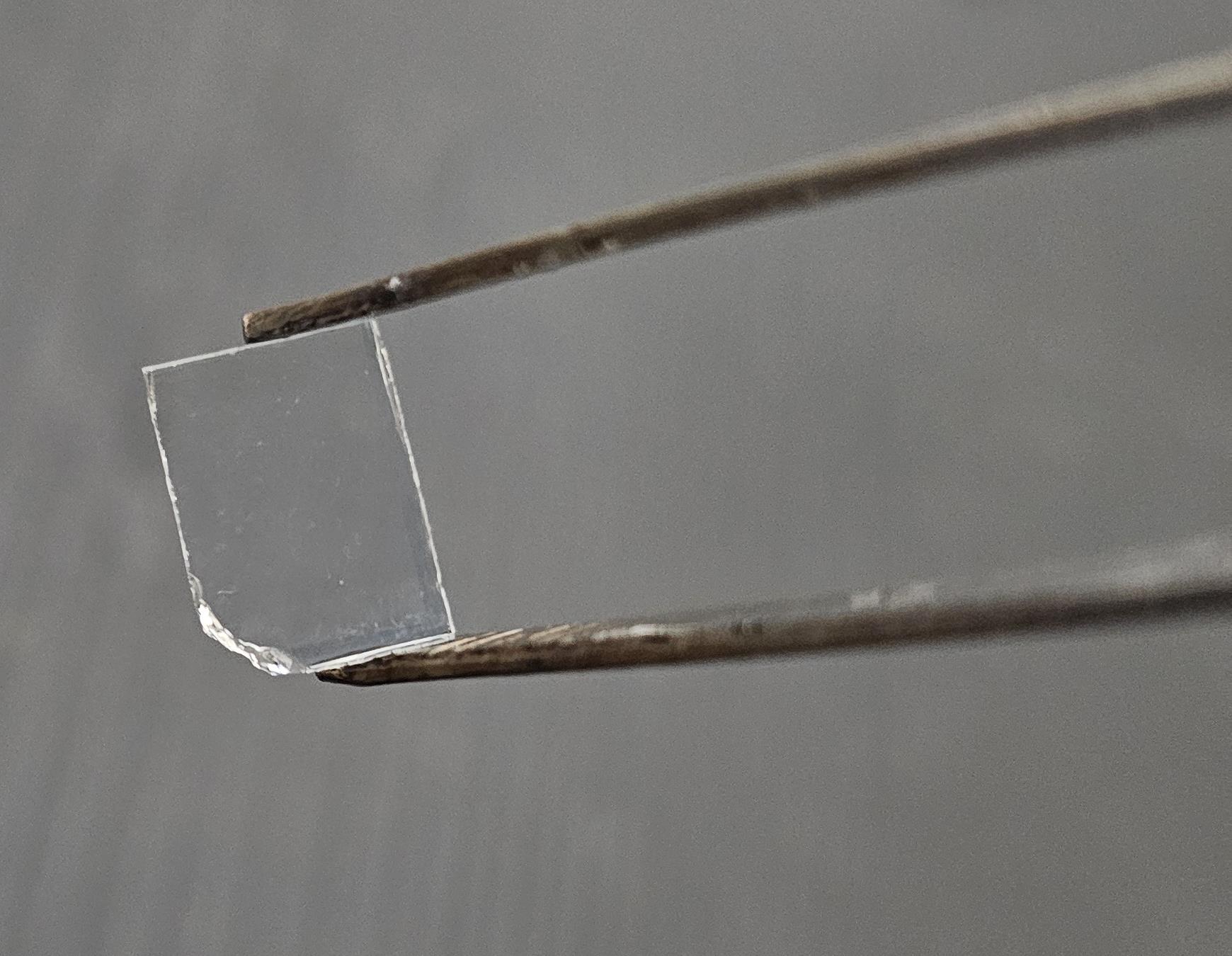
Figure 2. Slide piece with blunt end cut
4. Remove the glass debris generated during cutting and reclean the slide pieces with absolute ethanol to eliminate marker residues.
5. Wipe the cleaned slide pieces using lint-free tissue paper, wrap them, and sterilize them by autoclaving.
C. Slide embedding technique for fungal interaction studies
1. Prepare and sterilize PDA medium as described in steps A1–2.
2. Place sterile Petri dishes inside the laminar airflow chamber and mark a square corresponding to the dimensions of the glass slide piece on the bottom surface of each plate (Figure 3).
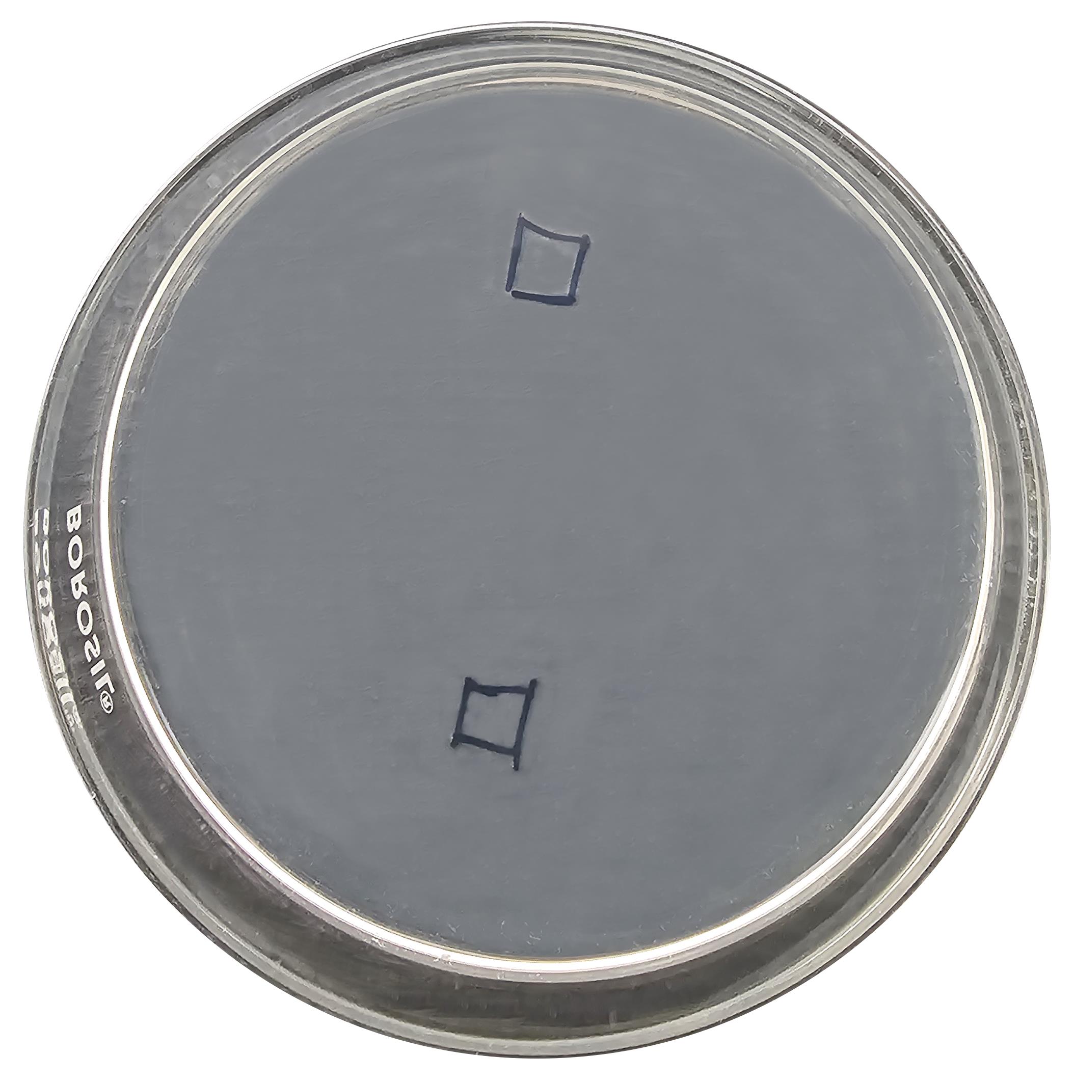
Figure 3. Petri plate marking. Marked regions indicate the positions for slide placement and fungal inoculation.
3. Pour molten PDA medium into the plates to a depth equal to the thickness of the slide piece (approximately 10 mL) and allow it to solidify.
4. Heat a sterile inoculation loop until red hot and aseptically remove a square section of agar corresponding to the marked dimensions from the solidified medium (Video 1).

5. Handle the autoclaved slide pieces using sterile forceps. Briefly dip the slide pieces in acetone, flame-sterilize them, and allow them to cool for 2 min.
Note: Acetone is used to remove residual grease and dust from the glass surface and serves as a combustion aid for subsequent flame sterilization.
Caution: Acetone is highly flammable. Keep it away from open flames except during the intended flame sterilization step. Handle acetone-soaked glass pieces with extreme care and follow appropriate fire safety precautions.
6. Carefully place the slide piece into the agar cavity created in the PDA plate (Figure 4A).
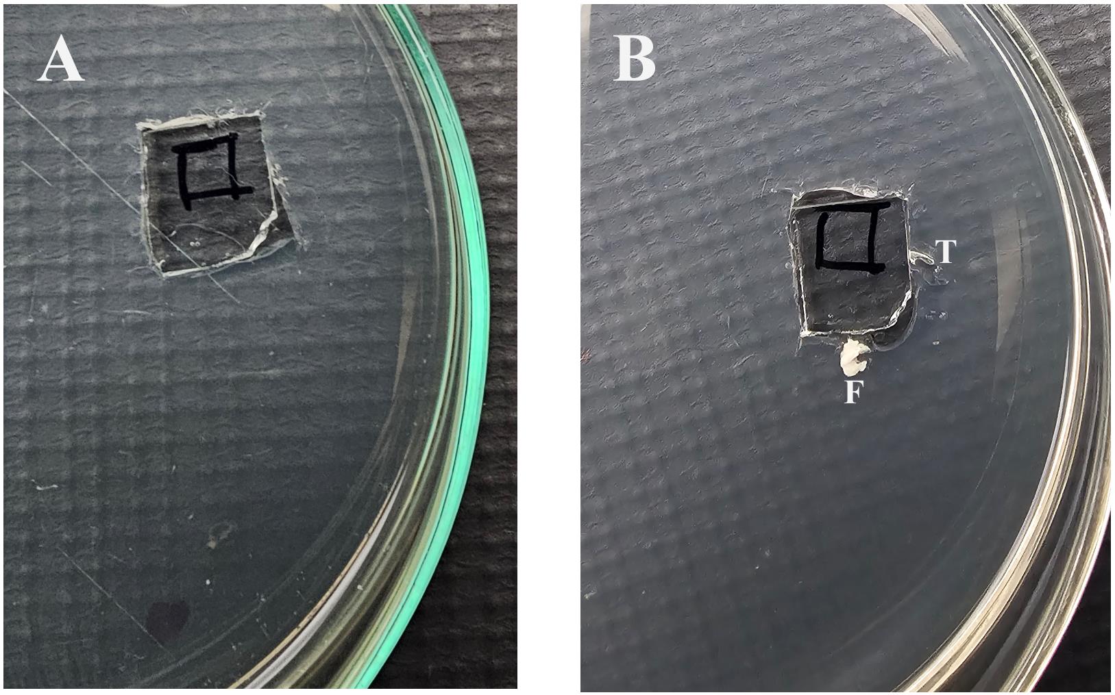
Figure 4. Slide piece placement and inoculation. (A) Slide piece placed on the agar surface. (B) Inoculation zones labeled for Fusarium sp. (F) and Trichoderma viride (T).
7. Inoculate the junction between the PDA medium and the embedded slide piece with a loopful of actively growing 7-day-old culture of Fusarium sp. (Video 2).
8. Seal the plates with parafilm and incubate them at 25 °C for 48 h.
Note: To obtain reproducible fungus–fungus interactions, the inoculation sequence, incubation period, and incubation temperature should be optimized according to the growth characteristics of the fungal species. The pathogen should be inoculated first and incubated under its optimal to sub-optimal growth conditions (temperature and duration) until it establishes sufficient growth along the PDA–glass interface, spreading bidirectionally from the inoculated edge, with the advancing mycelial front reaching approximately the midpoint of the two adjacent sides of the embedded slide piece. The fungal parasite should then be inoculated adjacent to the advancing hyphal margin of the pathogen (Figure 5). If the two fungi have different optimum growth temperatures, the incubation conditions should be adjusted accordingly following the second inoculation. In the present study, both the pathogen (Fusarium sp.) and the fungal parasite (Trichoderma viride) exhibited optimum growth at 25 °C; therefore, all incubations were performed at 25 °C. This optimization strategy ensures that the pathogen is well established without overgrowth while allowing the fungal parasite to grow toward the pathogen, thereby producing consistent and reproducible mycoparasitic interactions suitable for downstream SEM analysis. Because incubation temperature can influence fungal growth, host–parasite interactions, and the extent of mycoparasitism, it should be selected carefully according to the biological requirements of the interacting fungal species. After visible hyphal growth spreads across the slide surface, inoculate Trichoderma viride from a 7-day-old culture at the advancing margin of the Fusarium growth (Figure 4B).
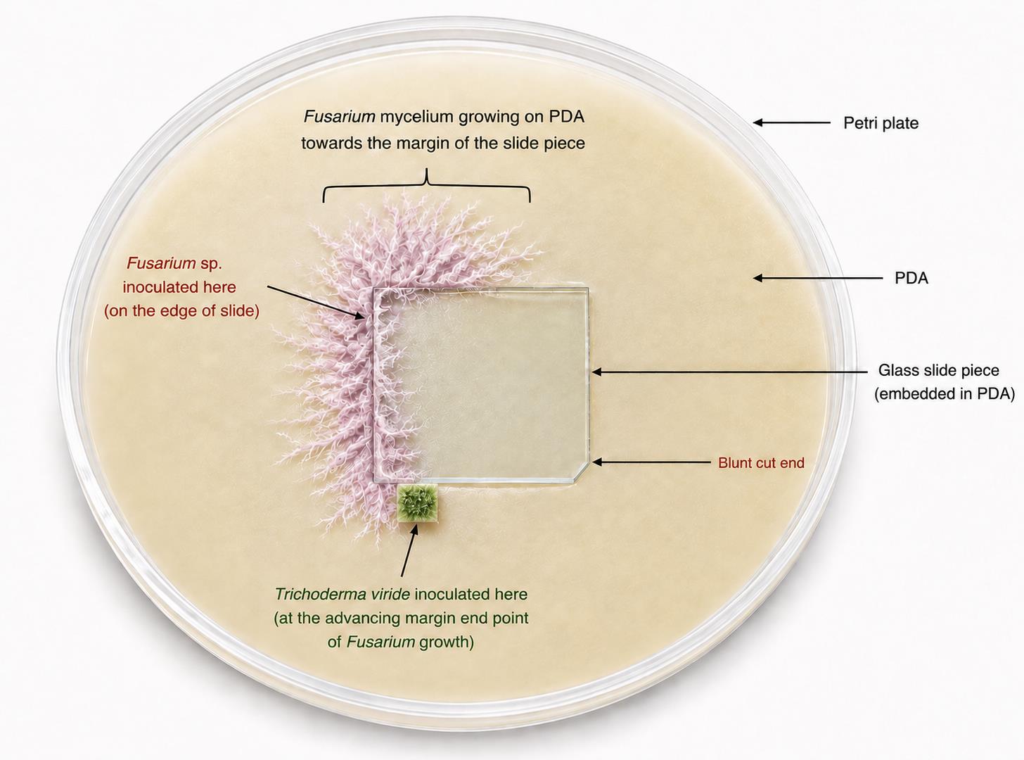
Figure 5. Schematic diagram of a 48-h-incubated Petri plate illustrating Fusarium sp. growth and the inoculation point of Trichoderma viride
9. Incubate the plates for an additional 48 h (Figure 6).
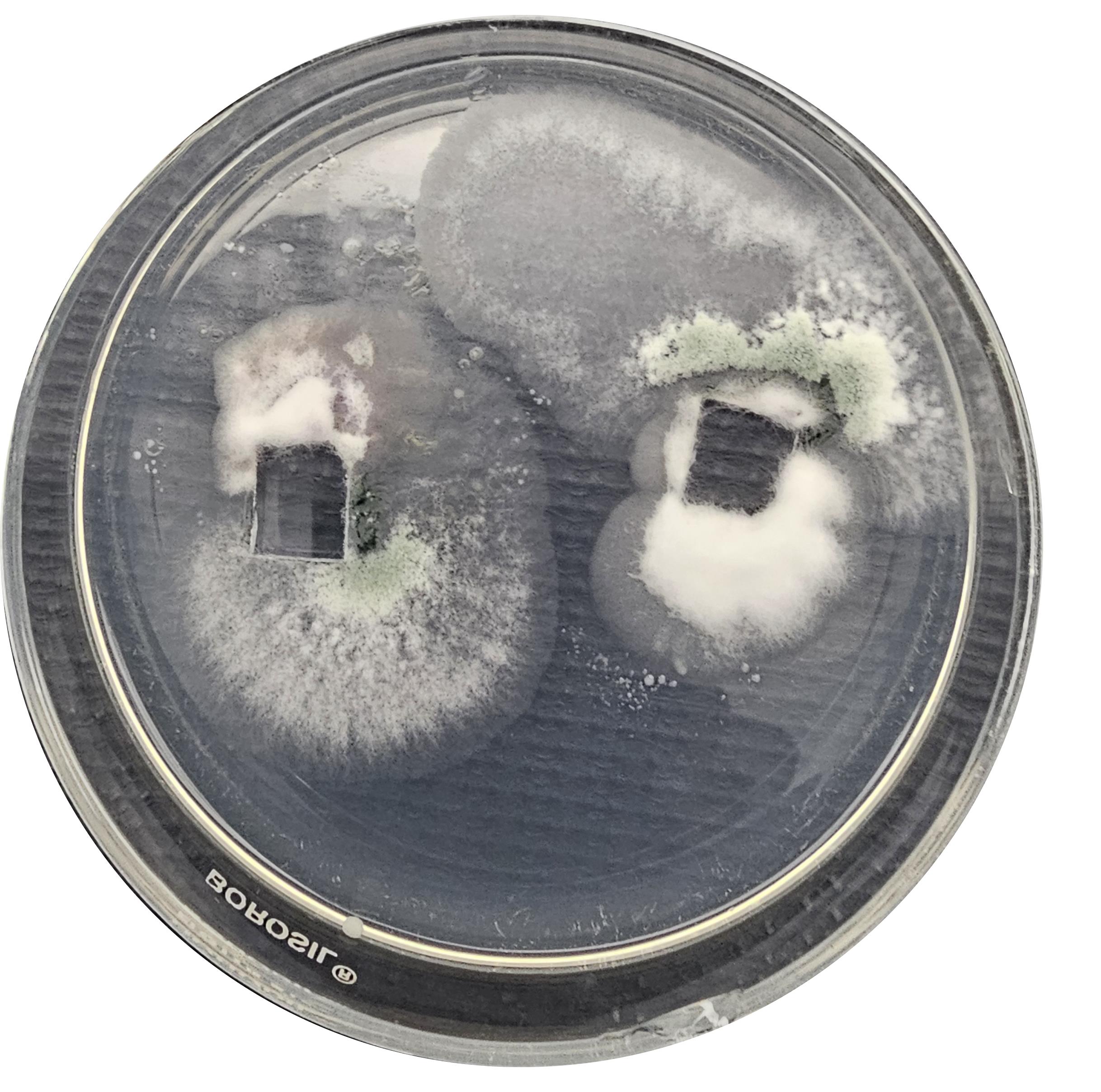
Figure 6. Fully grown fungal cultures around the glass slides
10. Examine the interaction zone on the slide piece under light to confirm fungal growth and interaction (Figure 7).
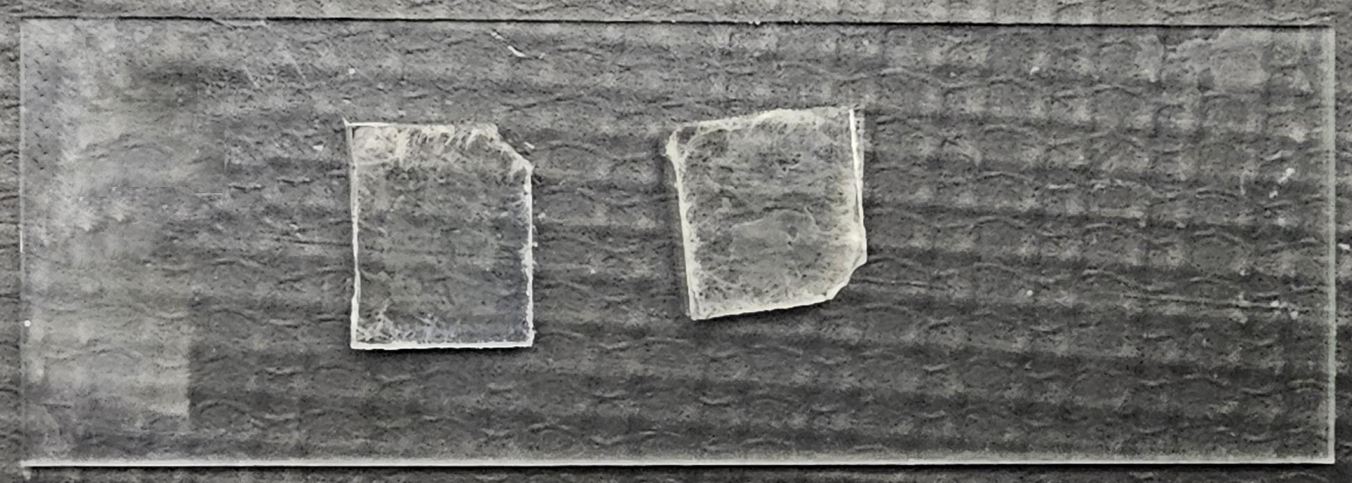
Figure 7. Slide pieces with fungal interaction
11. Carefully remove the slide pieces using sterile forceps and mount them onto clean, sterile glass slides for microscopic examination.
12. Identify the precise location of fungal interaction under a brightfield microscope and document it for subsequent SEM observation.
D. Fixation and dehydration of fungal interaction samples
1. Immediately after identifying the interaction zone, transfer the slide pieces along with the glass slide into a sterile Petri dish.
Critical: Transfer the slide pieces using forceps by firmly holding only the two opposite edges. Avoid bringing the forceps into contact with the interacting surface, as this may damage the specimen. Follow this precaution in all subsequent steps involving slide-piece transfer (Figure 2).
2. Using a Pasteur pipette, add 2–3 drops of glutaraldehyde fixative solution to completely cover the fungal interaction area on the slide pieces (Figure 8A).
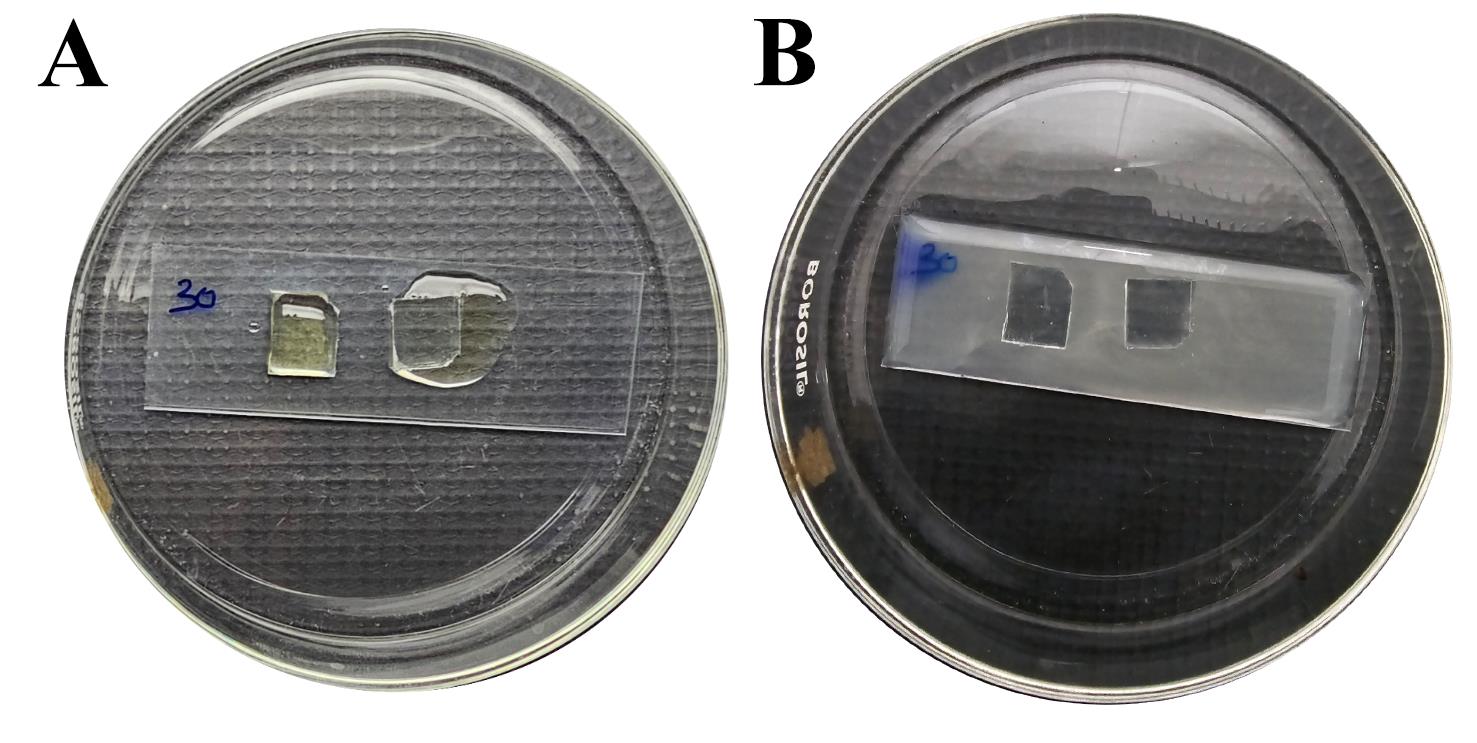
Figure 8. Fixation and dehydration. (A) Glutaraldehyde fixative covering the slide piece. (B) Washing and dehydration of the slide piece.
Critical: Due to the low volume of fixative, ensure that the solution does not dry during the subsequent incubation step. Alternatively, immerse the slide pieces completely in the fixative or add additional fixative to prevent drying.
3. Cover the Petri dish and incubate overnight (~14 h) at 4 °C in a refrigerator.
Critical: Do not incubate for more than 24 h.
Note: The fixation time and temperature may be adjusted depending on the sample and experimental requirements. While fixation for approximately 4 h at room temperature is generally sufficient, overnight fixation at 4 °C is recommended to achieve optimal preservation of fungal morphology and consistent SEM imaging outcomes.
4. Carefully discard the fixative solution without disturbing the slide pieces. Wash the slide pieces three times for 5 min each using 2–3 drops of sterile Milli-Q water to completely cover the surface.
Note: Perform washing by alternately transferring the slide pieces between two identical Petri dish setups containing glass slides. Transfer the slide pieces from the first setup to the second setup, add the respective reagent to the second setup, and then discard and renew the reagent in the first setup for the subsequent wash or treatment. Follow the same approach in the subsequent steps.
5. Perform dehydration sequentially using graded ethanol concentrations of 10%, 20%, 40%, 60%, 80%, and 100%.
6. Treat the samples thrice for 5 min each at every ethanol concentration by completely covering the slide surface with ethanol. Carry out the dehydration process in ascending order of ethanol concentration (Figure 8B).
Critical: Immediately proceed to the subsequent steps after 100% ethanol dehydration to minimize moisture exposure. Time is critical, but ensure that the ethanol has completely evaporated from the slide surface before proceeding to the next step.
7. Mount the dehydrated slide pieces carefully onto double-sided adhesive tape fixed inside a sterile 60 mm Petri dish to minimize disturbance during handling (Figure 9).
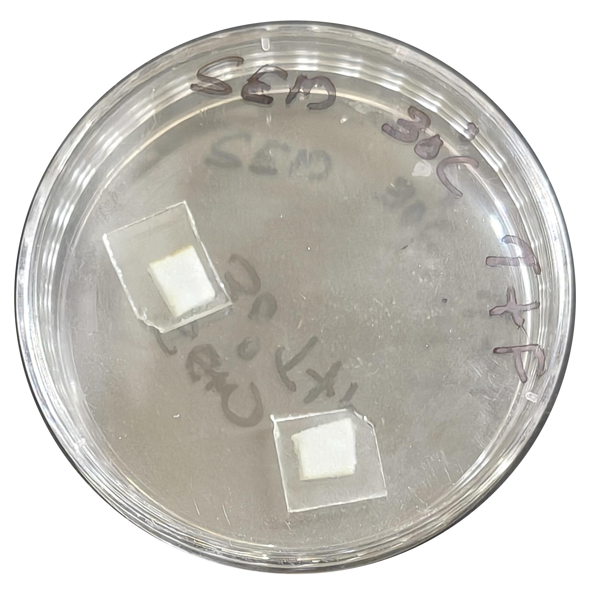
Figure 9. A 60-mm tissue dish with a slide piece attached to the double-sided adhesive tape
8. Seal the Petri dishes tightly with parafilm to prevent further moisture exposure.
9. Store the prepared samples in an incubator at 37 °C until SEM analysis or for up to 1 month.
E. Preparation and observation of samples under SEM
1. Mount the dehydrated slide pieces containing the fungal interaction zone onto SEM stubs using double-sided conductive adhesive tape prior to SEM analysis.
2. Sputter-coat the mounted samples with a thin (0.2–0.5 nm) gold-palladium conductive metal layer to enhance conductivity and improve image quality.
3. Observe the samples under a SEM using appropriate accelerating voltage (10–12 kV), working distance (10.5–19.5 mm), and magnification settings (1,500–4,000×) to visualize the interaction between Fusarium sp. and Trichoderma viride (Figure 10) (see Troubleshooting 2).
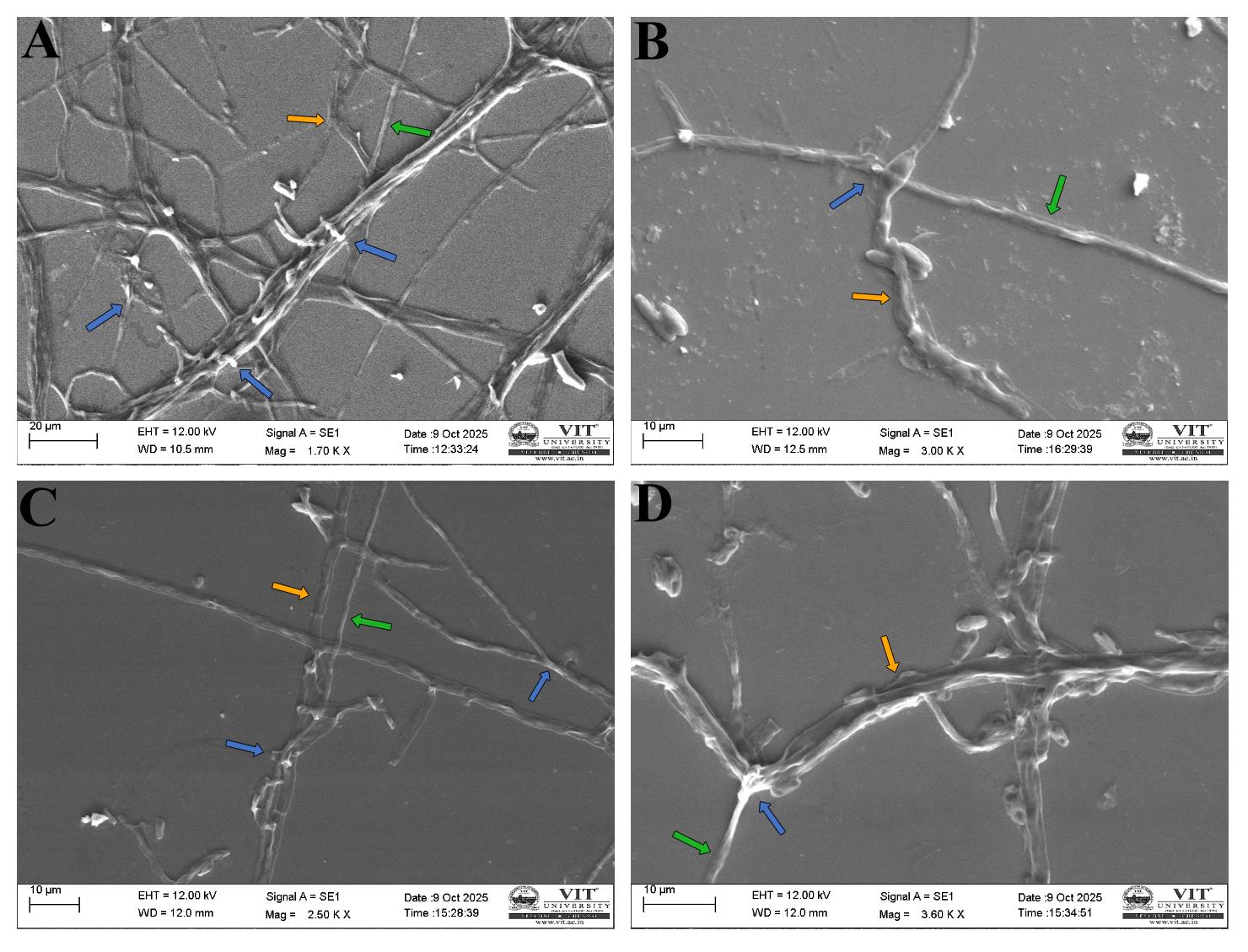
Figure 10. Scanning electron microscopy (SEM) images of interaction. Visible interaction between Trichoderma viride and Fusarium sp. Blue arrows indicate mycoparasitic interactions; green arrows indicate Trichoderma viride; orange arrows indicate Fusarium sp. (A) Coiling. (B) Cellular distortion. (C, D) Looping and cellular distortion.
Data analysis
During SEM acquisition, the blunt-end cut is used to locate the interaction zone between Trichoderma and Fusarium. The region is then appropriately magnified to identify specific interaction sites and acquire high-resolution images. Fusarium hyphae appear broader and larger (~2.98 ± 0.78 μm), whereas Trichoderma is observed as thin filamentous hyphae (~1.46 ± 0.53 μm). Hyphal coiling and looping of Trichoderma around Fusarium and cellular distortion of Fusarium hyphae are observed in Figure 9.
Validation of protocol
1. The protocol was validated using eight independent biological replicates to assess its reproducibility for visualizing mycoparasitic interactions between Fusarium sp. and Trichoderma viride by SEM.
2. In all replicates, Fusarium sp. was inoculated first and incubated for 48 h at 25 °C, followed by inoculation of T. viride and a further 48-h incubation at 25 °C. This sequential inoculation consistently produced intact pathogen hyphae and reproducible fungus–fungus interactions without pathogen overgrowth.
3. Some of the characteristic mycoparasitic interactions, including hyphal coiling, distortion, and degradation, were successfully observed in all eight biological replicates, demonstrating the reproducibility of the protocol.
4. An incubation period of 48 h for these fungi was found to be optimal. Longer incubation periods resulted in excessive mycelial growth, making the interaction zone overcrowded and more difficult to identify during SEM analysis.
5. To facilitate rapid localization of the interaction site during SEM, the interaction zone was first identified under a light microscope before fixation, and its position was recorded relative to the blunt-cut edge of the slide piece. This significantly reduced the time required to locate the interaction during SEM imaging.
6. The blunt-cut edge also served as a permanent orientation marker during fixation, dehydration, storage, and transportation, preventing accidental inversion of the slide pieces and ensuring that the interaction surface remained correctly oriented.
7. Throughout all these biological replicates, the prepared slide pieces remained intact during handling, storage, and transportation, enabling successful SEM imaging without damage or loss of the fungal interaction region.
General notes and troubleshooting
General notes
1. Maintain a dust-free working environment throughout the experiment to minimize contamination and artifact formation.
2. All fungal culture handling should preferably be performed inside a laminar airflow chamber to prevent spore dispersal and ensure aseptic conditions.
3. This technique can be used for fungal morphology visualization under SEM, other than interaction studies.
4. During incubation, fungal cultures should be monitored carefully to prevent overgrowth, which may result in dense filamentous structures and a crowded slide appearance that interferes with microscopic observation.
Troubleshooting
Problem 1: Glutaraldehyde fixative precipitation.
Possible causes: Improper temperature or pH.
Solution: Glutaraldehyde prepared in phosphate buffer may precipitate at room temperature (approximately 25–30 °C). To avoid precipitation, the glutaraldehyde fixative solution should be freshly prepared and used immediately.
Problem 2: Bright patches on the SEM field.
Possible causes: Exposure to moisture or improper drying during storage.
Solution: Excessive white appearance or glare in filamentous fungal samples may indicate incomplete drying after the dehydration process or exposure of the sample to moisture/humidity. To prevent such artifacts, ensure that the smears are completely dried before SEM analysis. Additionally, seal the SEM slides tightly with parafilm during storage and transportation, and preferably store them under warm conditions (≥37 °C) to minimize exposure to atmospheric moisture and humidity.
Problem 3: Excessive or insufficient fungal growth before interaction.
Possible cause: Incubation period not optimized according to the growth rate of the fungal isolates.
Solution: Adequate pathogen growth is achieved when the advancing mycelial front reaches the adjacent margins of the embedded slide piece. If the pathogen has not reached these margins, extend the incubation period before inoculating the fungal parasite. If the pathogen extends beyond the intended interaction zone or the slide surface becomes overcrowded with mycelia, reduce the incubation period before the second inoculation. The fungal parasite should be inoculated immediately adjacent to the advancing margin of the pathogen to ensure reproducible interactions and facilitate SEM visualization.
Acknowledgments
The authors are grateful to the Vellore Institute of Technology, Vellore, Tamil Nadu, India, for providing the facilities, support, and encouragement. This work was supported by the seed grant provided by Vellore Institute of Technology, Vellore, India (sanction order no. SG20230036). S.P. acknowledges RV University, Mysuru, for infrastructural support and encouragement. The authors also acknowledge Elamala Biotech Lab for kindly providing fungal cultures and infrastructural support. The authors acknowledge the use of AI-assisted tools (Figurelabs) to generate the Graphical overview and Figure 5 in this manuscript. All images were critically evaluated and verified for scientific accuracy by the authors, who take full responsibility for the final content.
Author contributions
Conceptualization, S.P., S.T.; Investigation, S.T.; Writing—Original Draft, S.T.; Writing—Review & Editing, S.P.; Funding acquisition, S.P.; Supervision, S.P.
Competing interests
Dr. Shweta Panchal is an Associate Editor for Bio-protocol but did not participate in the editorial and peer review process of this article, except as an author. The authors declare no other conflict of interest.
References
- Viterbo, A. and Horwitz, B. A. (2014). Mycoparasitism. In K. A. Borkovich and D. J. Ebbole (Eds.), Cellular and Molecular Biology of Filamentous Fungi (pp. 676–693). Washington, DC, USA: ASM Press. https://doi.org/10.1128/9781555816636.ch42
- Jeffries, P. (1995). Biology and ecology of mycoparasitism. Can J Bot. 73: 1284–1290. https://doi.org/10.1139/b95-389
- Sun, J. Z., Liu, X. Z., McKenzie, E. H. C., Jeewon, R., Liu, J. K., Zhang, X. L., Zhao, Q. and Hyde, K. D. (2019). Fungicolous fungi: terminology, diversity, distribution, evolution, and species checklist. Fungal Diversity. 95(1): 337–430. https://doi.org/10.1007/s13225-019-00422-9
- Atanasova, L., Crom, S. L., Gruber, S., Coulpier, F., Seidl-Seiboth, V., Kubicek, C. P. and Druzhinina, I. S. (2013). Comparative transcriptomics reveals different strategies of Trichodermamycoparasitism. BMC Genomics. 14(1): e1186/1471–2164–14–121. https://doi.org/10.1186/1471-2164-14-121
- Mukherjee, P. K., Mendoza-Mendoza, A., Zeilinger, S. and Horwitz, B. A. (2022). Mycoparasitism as a mechanism of Trichoderma-mediated suppression of plant diseases. Fungal Biol Rev. 39: 15–33. https://doi.org/10.1016/j.fbr.2021.11.004
- Bozzola, J. J. and Russell, L. D. (2006). Electron microscopy: principles and techniques for biologists (2. ed., [Nachdr.]). Boston: Jones and Bartlett.
- Brundrett, M., Bougher, N., Dell, B., Grove, T. and Malajczuk, N. (1996). Working with Mycorrhizas in Forestry and Agriculture. Australian Centre for International Agricultural Research. https://doi.org/10.13140/2.1.4880.5444
- Riddell, R. W. (1950). Permanent Stained Mycological Preparations Obtained by Slide Culture. Mycologia. 42(2): 265–270. https://doi.org/10.1080/00275514.1950.12017830
- Bhat, K. A. (2017). A New Agar Plate Assisted Slide Culture Technique to Study Mycoparasitism of Trichoderma sp. on Rhizoctonia solani and Fusarium oxysporium. Int J Curr Microbiol Appl Sci. 6(8): 3176–3180. https://doi.org/10.20546/ijcmas.2017.608.378
Article Information
Publication history
Received: May 14, 2026
Accepted: Jul 16, 2026
Available online: Aug 14, 2026
Published: Sep 5, 2026
Copyright
© 2026 The Author(s); This is an open access article under the CC BY-NC license (https://creativecommons.org/licenses/by-nc/4.0/).
How to cite
Tomichen, S. and Panchal, S. (2026). A Modified Slide-Embedded Scanning Electron Microscopy Preparation Method to Visualize Antagonistic Interactions Between Trichoderma viride and Fusarium sp.. Bio-protocol 16(17): e5805. DOI: 10.21769/BioProtoc.5805.

 Download Citation in RIS Format
Category
Microbiology > Microbial cell biology > Cell imaging
Microbiology > Microbe-host interactions > Fungus
Environmental science > Fungus
Do you have any questions about this protocol?
Post your question to gather feedback from the community. We will also invite the authors of this article to respond.
 Post a Question


