


- Protocols

- Articles and Issues
- For Authors
- About
- Become a Reviewer


Evaluating Thioredoxin-Mediated CFoCF1 Reduction Using an In Vitro Thylakoid Assay

Published: Vol 16, Iss 11, Jun 5, 2026 DOI: 10.21769/BioProtoc.5706 Views: 357
Reviewed by: Felix BuchertAnonymous reviewer(s)
 PDF
PDF  Ask a question
Original research article
The authors used this protocol in:
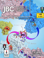
Sep 2024
Advertisement



Protocol Collections
Comprehensive collections of detailed, peer-reviewed protocols focusing on specific topics




Abstract
The activity of chloroplast ATP synthase (CFoCF1) is precisely regulated through a thioredoxin (Trx)-mediated dithiol/disulfide reaction in response to varying light conditions. This regulatory mechanism is further controlled by ΔpH formation across the thylakoid membrane. To better understand this complicating regulatory function of CFoCF1, a method is required to evaluate the extent of CFoCF1 reduction by Trx under controlled ΔpH conditions and to directly evaluate the redox state of CFoCF1. In this study, we present a simple in vitro procedure to assess the CFoCF1 reduction system using spinach thylakoids. The method consists of three key steps: (A) simple preparation of intact thylakoids from spinach leaves; (B) reduction of CFoCF1 on the thylakoid membrane using recombinant Trx under light irradiation; and (C) in situ determination of the redox state of CFoCF1 by labeling thiol groups with a maleimide reagent followed by protein detection using western blotting. The redox state of CFoCF1 was determined by mobility shifts on non-reducing SDS-PAGE. This protocol provides a refined strategy for elucidating the regulatory mechanism controlling energy conversion by CFoCF1 under fluctuating photosynthetic conditions.
Key features
• A simple isolation method for intact thylakoids from Spinacia oleracea that enables the evaluation of the reduction of chloroplast ATP synthase by thioredoxin.
• The combination of LED light irradiation and electron mediators allows controlled adjustment of the proton electrochemical gradient across thylakoid membranes.
• The redox state of chloroplast ATP synthase can be distinguished by labeling free thiols with maleimide reagents and quantitative detection by western blotting.
Keywords: Chloroplast ATP synthase Thioredoxin
Thylakoid membrane
Redox regulation
Thiol labeling
Western blotting
Spinacia oleracea
Thioredoxin
Thylakoid membrane
Redox regulation
Thiol labeling
Western blotting
Spinacia oleracea
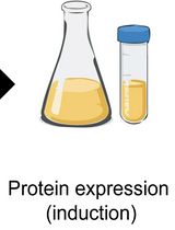
Graphical overview
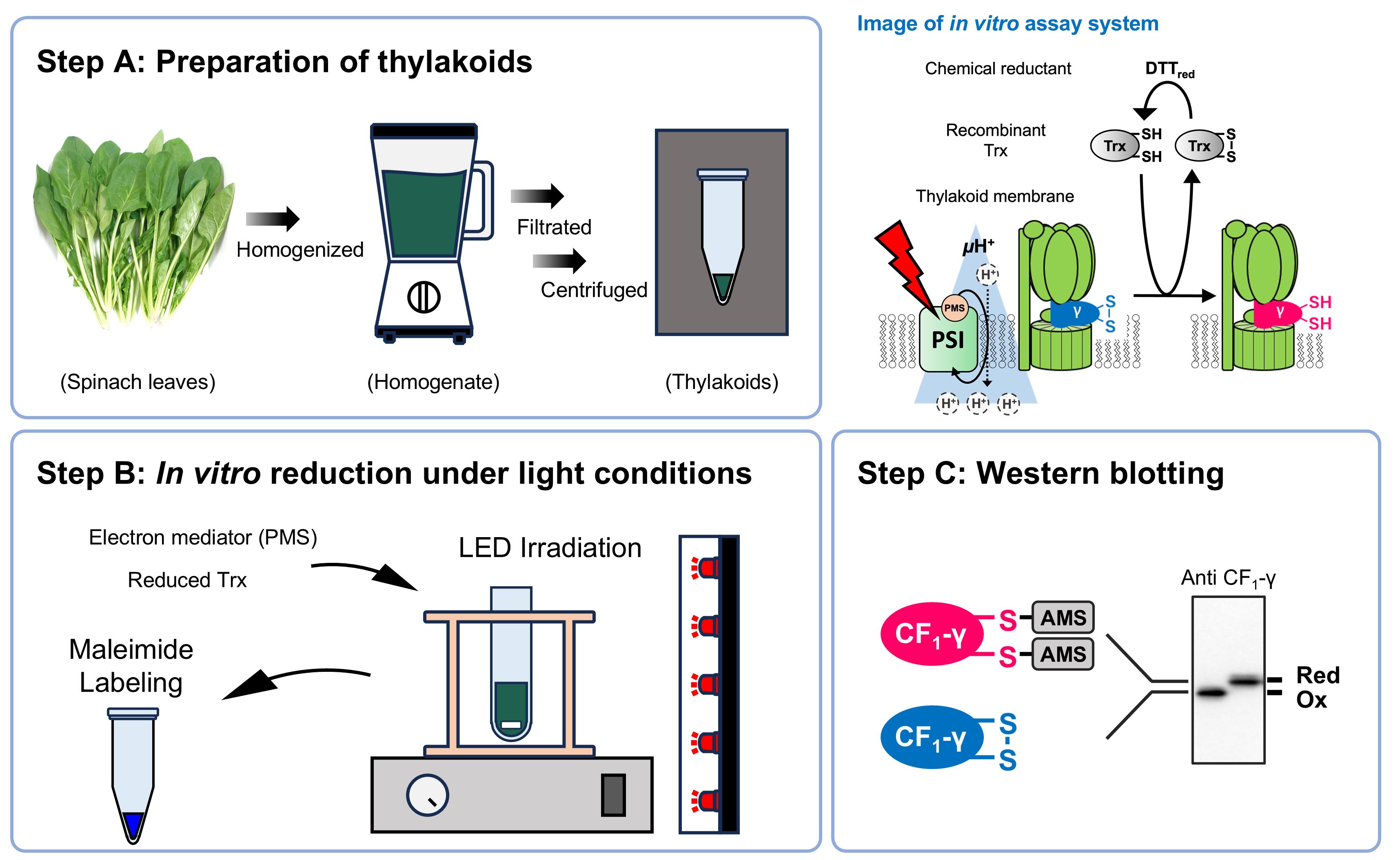
Background
Chloroplast ATP synthase (CFoCF1) is a pivotal enzyme in photosynthetic energy conversion in green plants. When photosynthetic electron transport generates a proton electrochemical gradient (ΔμH+) across the thylakoid membrane under light conditions, this gradient is primarily consumed by CFoCF1 to drive ATP synthesis [1–3]. Therefore, CFoCF1 activity is strongly regulated to maintain efficient energy conversion under varying light conditions. One representative regulatory mechanism is redox control [4,5]. The γ subunit (CF1-γ), which rotates within the enzyme complex during catalysis, harbors a redox-active cysteine pair [6,7]. These cysteines are targets of thioredoxin (Trx), which mediates the reducing power from the photosynthetic electron transport chain [8]. Furthermore, CF1-γ is reduced by Trx only when ΔpH is formed across the thylakoid membrane under photosynthetic conditions [9–11]. Thus, CFoCF1 is regulated through a multistep control mechanism involving Trx-mediated redox reactions, closely linked to ΔpH formation, reflecting changes in light environment.
Over the past half-century of research on CF1, including by our research group, methods for examining the redox state of CF1-γ have been established. Jagendorf first demonstrated ATP synthesis driven by ΔpH across thylakoid membranes in isolated chloroplasts using the so-called acid–base transition experiment, in which chloroplasts were sequentially incubated in buffers of different pHs [12]. Subsequently, Gräber et al. applied the acid–base transition method to isolated thylakoids or liposomes containing isolated CFoCF1 in the presence of excess reducing agents, enabling kinetic analysis of the relationship between the redox state of CF1-γ and CFoCF1 enzymatic activity [13,14]. In another approach, Schwarz et al. investigated the selectivity of Trx toward CF1-γ by measuring ATP hydrolysis activity under defined pH gradients across thylakoid membranes in the presence of different Trx isoforms [15]. However, these studies focused primarily on the enzymatic activity of CFoCF1 rather than directly determining the exact redox state of CF1-γ. Because the extent of ΔpH affects both ATP synthesis and hydrolysis activities of CFoCF1, a system capable of directly monitoring the redox state of CF1-γ under ΔpH-forming conditions, independent of enzyme activity measurements, is required. In previous work, our laboratory successfully determined the redox state of CF1-γ by directly applying a maleimide reagent to isolated spinach chloroplasts under light conditions [16]. Using this approach, we observed that CF1-γ is rapidly reduced upon light irradiation, and that reduction is completely inhibited in the presence of an uncoupler in the chloroplast solutions.
In this study, we refine this conventional approach and provide a simple protocol for quantitative reduction analysis of CF1-γ using isolated spinach thylakoids combined with an artificial electron transfer mediator, which is required for membrane potential formation in the light [17], and recombinant Trx. Using this method, we successfully demonstrated the selectivity of major Trx family proteins toward CF1-γ [18], oxidation of CF1-γ by Trx-like proteins associated with ΔpH dissipation [19], and the distinct contributions of ΔpH and membrane potential to CF1-γ reduction [20], supporting the sensitivity and accuracy of this method.
Materials and reagents
Biological materials
1. Fresh bunched spinach (Spinacia oleracea) (purchased from the market)
Reagents
1. Distilled water
2. Tricine (Wako, catalog number: 347-02844)
3. Magnesium chloride hexahydrate (MgCl2·6H2O) (Nacalai Tesque, catalog number: 20909-55)
4. Sodium chloride (NaCl) (Nacalai Tesque, catalog number: 31320-05)
5. Potassium chloride (KCl) (Nacalai Tesque, catalog number: 28513-85)
6. Sodium hydroxide (NaOH) (Nacalai Tesque, catalog number: 31511-05)
7. Sucrose (Nacalai Tesque, catalog number: 30404-45)
8. Dithiothreitol (DTT) (Wako, catalog number: 049-08972)
9. 1-methoxy-5-methylphenazinium methylsulfate (1-Methoxy PMS) (Sigma-Aldrich, catalog number: M8640)
10. Carbonyl cyanide 4-(trifluoromethoxy)phenylhydrazone (FCCP) (Sigma-Aldrich, catalog number: C2920)
11. Ethanol (Nacalai Tesque, catalog number: 14713-24)
12. Sodium lauryl sulfate (SDS) (Nacalai Tesque, catalog number: 31607-65)
13. Glycerol (Nacalai Tesque, catalog number: 17018-83)
14. Bromophenol blue (BPB) (Wako, catalog number: 029-02912)
15. 4-acetamido-4’-maleimidylstilbene-2,2’-disulfonate (AMS) (Invitrogen, catalog number: A485)
16. Tris(hydroxymethyl)aminomethane (Tris) (Nacalai Tesque, catalog number: 35434-21)
17. Glycine (Nacalai Tesque, catalog number: 17141-95)
18. Polyoxyethylene (20) sorbitan monolaurate (Tween 20) (Wako, catalog number: 167-11515)
19. Methanol (Nacalai Tesque, catalog number: 21914-74)
20. Disodium hydrogen phosphate (Nacalai Tesque, catalog number: 31801-05)
21. Potassium dihydrogen phosphate (Nacalai Tesque, catalog number: 28721-55)
22. SDS-PAGE protein marker (Bio-Rad, catalog number: 161-0373)
23. ECL Prime western blotting detection reagent (GE Healthcare, catalog number: RPN2232)
24. Primary antibody for the protein of interest
25. Secondary antibody for the protein of interest
26. Skim milk (Morinaga Nyugyo, catalog number: 0652842)
27. 100% (w/v) trichloroacetic acid (TCA) (Wako, catalog number: 200-08085)
28. Acetone (Wako, catalog number: 019-00353)
Solutions
1. 4× stock solution for grinding buffer (see Recipes)
2. Grinding buffer (see Recipes)
3. 25× DTT stock solution (see Recipes)
4. 50× PMS stock solution (see Recipes)
5. FCCP stock solution (see Recipes)
6. Thiol-labeling solution (see Recipes)
7. Electrophoresis buffer for SDS-PAGE (see Recipes)
8. Transfer buffer for western blotting (see Recipes)
9. PBS buffer (see Recipes)
Recipes
1. 4× stock solution for grinding buffer
| Reagent | Final concentration | Quantity or volume |
|---|---|---|
| Tricine | 200 mM | 35.8 g |
| MgCl2·6H2O | 20 mM | 4.06 g |
| NaCl | 40 mM | 2.34 g |
| KCl | 200 mM | 14.9 g |
| Distilled water | N/A | Up to 1 L |
| Total | N/A | 1 L |
Adjust pH to 7.5 using NaOH. Prepare in a media bottle and store at 4 °C in a refrigerator; it is stable for several months.
2. Grinding buffer
| Reagent | Final concentration | Quantity or volume |
|---|---|---|
| 4× stock solution for grinding buffer | N/A | 100 mL |
| Sucrose | 0.4 M | 54.8 g |
| Distilled water | N/A | Up to 400 mL |
| Total | N/A | 400 mL |
Prepare in a media bottle. Always use a freshly prepared grinding buffer for each experiment and maintain it at low temperature until just before use.
3. 25× DTT stock solution
| Reagent | Final concentration | Quantity or volume |
|---|---|---|
| DTT | 500 mM | 77.1 mg |
| Distilled water | N/A | 1 mL |
Prepare in a microcentrifuge tube and store at -20 °C in a freezer; stable for several months. Dilute the 25× DTT stock solution to 1× (2 mM DTT) with grinding buffer before use.
4. 50× PMS stock solution
| Reagent | Final concentration | Quantity or volume |
|---|---|---|
| 1-Methoxy PMS | 10 mM | 3.36 mg |
| Distilled water | N/A | 1 mL |
Prepare in a microcentrifuge tube and store in a storage box away from light at room temperature; stable for several months. Dilute the 50× PMS stock solution to 1× (200 μM 1-Methoxy PMS) with grinding buffer before use.
5. FCCP solution
| Reagent | Final concentration | Quantity or volume |
|---|---|---|
| FCCP | 2 mM | 1.02 mg |
| Ethanol | N/A | 2 mL |
Prepare in a microcentrifuge tube and store at -20 °C in a freezer; stable for several months.
6. Thiol-labeling solution
| Reagent | Final concentration | Quantity or volume |
|---|---|---|
| Tris | 62.5 mM | 75.7 mg |
| SDS | 2% (w/v) | 200 mg |
| Glycerol | 7.5% (w/v) | 750 mg |
| BPB | 0.01% (w/v) | 1 mg |
| AMS | 2 mM | 10 mg |
| Distilled water | N/A | Up to 10 mL |
| Total | N/A | 10 mL |
Adjust pH to 6.8 using HCl. Prepare in a conical tube. Always use a freshly prepared thiol-labeling solution for each experiment. Store in a storage box away from light at room temperature until just before use.
7. Electrophoresis buffer for SDS-PAGE
| Reagent | Final concentration | Quantity or volume |
|---|---|---|
| Tris | 25 mM | 30.25 g |
| Glycine | 192 mM | 144.1 g |
| SDS | 0.1% (w/v) | 10 g |
| Distilled water | N/A | Up to 10 L |
| Total | N/A | 10 L |
Prepare in a solution tank and store at room temperature; stable for several months.
8. Transfer buffer for western blotting
| Reagent | Final concentration | Quantity or volume |
|---|---|---|
| Tris | 40 mM | 24.2 g |
| Glycine | 242 mM | 90.7 g |
| SDS | 0.1% (w/v) | 5 g |
| Ethanol | 20% (v/v) | 1 L |
| Distilled water | N/A | Up to 5 L |
| Total | N/A | 5 L |
Prepare in a solution tank and store at room temperature; stable for several months.
9. PBS buffer
| Reagent | Final concentration | Quantity or volume |
|---|---|---|
| NaCl | 137 mM | 80 g |
| KCl | 2.69 mM | 2 g |
| Na2HPO4 | 7.75 mM | 11 g |
| KH2PO4 | 1.47 mM | 2 g |
| Tween 20 | 0.4% (v/v) | 20 mL |
| Distilled water | N/A | Up to 10 L |
| Total | N/A | 10 L |
Prepare in a solution tank and store at room temperature; stable for several months.
Laboratory supplies
1. Newspaper
2. 1.5 mL microcentrifuge tube (WATSON, catalog number: 131-7155C)
3. 10 μL pipette tips (WATSON, catalog number: 110-201C)
4. 200 μL pipette tips (WATSON, catalog number: 110-705C)
5. 1,000 μL pipette tips (WATSON, catalog number: 110-804C)
6. Gauze (Iwatsuki, catalog number: 002-21540)
7. Absorbent paper (ATTO, catalog number: CB-09A)
8. Immun-Blot PVDF membrane (Bio-Rad, catalog number: 1620177)
Equipment
1. 10 μL pipette (Gilson, model: P10, catalog number: F144802)
2. 20 μL pipette (Gilson, model: P20, catalog number: F123600)
3. 200 μL pipette (Gilson, model: P200, catalog number: F123601)
4. 1,000 μL pipette (Gilson, model: P1000, catalog number: F123602)
5. Blender (Kuvings, model: KPB-351SP)
6. High-speed refrigerated centrifuge (Himac, model: CR20GIII)
7. Refrigerated microcentrifuge (TOMY, model: MX-307)
8. Equipment for SDS-PAGE (NIHON EIDO, model: NA-1012)
9. Equipment for western blotting (BIO CRAFT, model: BE-320)
10. Luminescence image analyzer (Fujifilm, model: LAS-3000 mini)
11. Spectrofluorometer (Jasco, model: FP-6500)
12. Quartz cell
13. Paintbrush
14. LED light (CCS Inc., model: ISLM-150150-HWHR)
15. Grass tube (AS ONE, catalog number: 14101005B)
16. Conical beaker (AS ONE, catalog number: 1-7117-03)
17. Funnel (AS ONE, catalog number: 6-319-06)
18. Magnetic stirrer (TOMY, model: FS-210)
Software and datasets
1. ImageJ, http://imagej.nih.gov/ij//
Procedure
A. Preparation of thylakoids from spinach leaves
1. Prepare grinding buffer (see Recipes) and keep it at 4 °C.
2. Obtain a bunch of spinach from a local supermarket one day before use.
3. Wash thoroughly with water, wrap in newspaper, and store overnight in the dark at 4 °C to deplete starch levels and improve the intactness of isolated thylakoids.
4. Excise approximately 10 g (fresh weight) of leaves (excluding petioles) in the dark.
5. Homogenize in 200 mL of grinding buffer using a blender (thrice for 3 s).
6. Filter through four layers of gauze using a funnel and collect the flowthrough.
7. Centrifuge at 3,000× g for 10 min at 4 °C.
8. Discard the supernatant and gently resuspend the pellet in 40 mL of grinding buffer on ice using a paintbrush.
9. Centrifuge at 300× g for 1 min at 4 °C, collect the supernatant, and centrifuge again at 3,000× g for 10 min at 4 °C.
10. Repeat steps A8–9 once more and resuspend the resulting pellet in 2 mL of grinding buffer using a paintbrush.
11. Dilute a 10 μL aliquot of the preparation solution 20-fold with the grinding buffer (e.g., 190 μL) to determine chlorophyll concentration.
12. Add 800 μL of 100% acetone to obtain 80% (v/v) aqueous acetone for extraction and vortex for 10 min.
13. Centrifuge at 20,400× g for 5 min at room temperature.
14. Transfer the supernatant to a quartz cell and measure absorbance at 646.6, 663.6, and 750 nm using a spectrophotometer.
15. Calculate the total concentration of chlorophyll (a + b) (mg/mL) using the following formula [21]:
[Chlorophyll a + b] = 17.76 × (Abs646.6 - Abs750) + 7.34 × (Abs663.6 - Abs750)
Note: The chlorophyll concentration measured here is a 100-fold diluted value from the original extract.
16. Keep the preparation on ice in the dark for at least 1 h before the assay.
17. Before use, dilute the preparation to a chlorophyll concentration (Chl) of 200 μg/mL.
B. In vitro assay of Trx-mediated CFoCF1 reduction
1. Use grinding buffer as the reaction medium for the in vitro reduction assay; perform the assay at 25 °C.
2. Incubate 100 μL of 20 μM Trx-f with 100 μL of 2 mM DTT in a tube for 5 min at 25 °C.
3. Add 200 μL of the Trx-f/DTT mixture and 50 μL of 200 μg Chl/mL thylakoids to 1,730 μL of grinding buffer in a glass tube and stir gently for 1 min using a magnetic stirrer.
4. Add 20 μL of 200 μM 1-Methoxy PMS and stir for 1 min.
5. Add 1 μL of 2 mM FCCP as a negative control when required.
6. Final concentrations in the mixture are 5 μg Chl/mL thylakoids, 2 μM 1-Methoxy PMS, 100 μM DTT, and 1 μM Trx-f.
7. Irradiate this mixture horizontally with red light at 660 nm for 5 min using an LED to initiate ΔμH+ formation and Trx-f-mediated reduction.
8. Continue stirring during the reaction and collect 100 μL aliquots at planned time points.
9. Immediately add 10% (w/v) TCA to stop the reduction by protein precipitation.
C. Determination of the protein redox state
This section is performed following step B of the procedure described in [22], with the following slight modifications:
1. Prepare a 10% polyacrylamide gel for SDS-PAGE, typically 85 × 60 × 1 mm.
2. Set the polyacrylamide gel in the electrophoresis tank and add the electrophoresis buffer.
3. Load 10 μL of each protein sample and run the electrophoresis.
4. Prepare the PVDF membrane and absorbent papers for western blotting. Hydrophilically treat the PVDF membrane with methanol (several seconds) and then equilibrate in the transfer buffer (for several minutes; see Recipes) prior to use.
5. Set the polyacrylamide gel after SDS-PAGE, the PVDF membrane, and the absorbent papers in the blotting equipment. Run the blotting.
6. After blotting, wash the PVDF membrane with distilled water and then PBS buffer (see Recipes) for several minutes.
7. Incubate the PVDF membrane with 1% (w/v) skim milk dissolved in PBS buffer overnight at 4 °C.
8. Incubate the PVDF membrane with the primary antibody diluted with PBS buffer at room temperature for 2 h.
9. Wash the PVDF membrane with PBS buffer thrice for 10 min.
10. Incubate the PVDF membrane with the secondary antibody diluted with PBS buffer at room temperature for 1 h.
11. Wash the PVDF membrane with PBS buffer thrice for 10 min.
12. Incubate the PVDF membrane with the ECL Prime solution and detect luminescence using a LAS-3000 Mini Imaging System.
Data analysis
1. The thiol-labeling maleimide reagent AMS (molecular mass 536.44) decreases protein mobility on SDS-PAGE (Figure 1A). The redox state of CF1-γ is identified by an observable band shift. Protein reduction levels are calculated by quantifying signal intensities of reduced and oxidized forms using ImageJ software (Figure 1B).
2. Sequential sampling under changing environmental conditions (e.g., light) enables tracking of protein redox behaviors under environmental fluctuations.
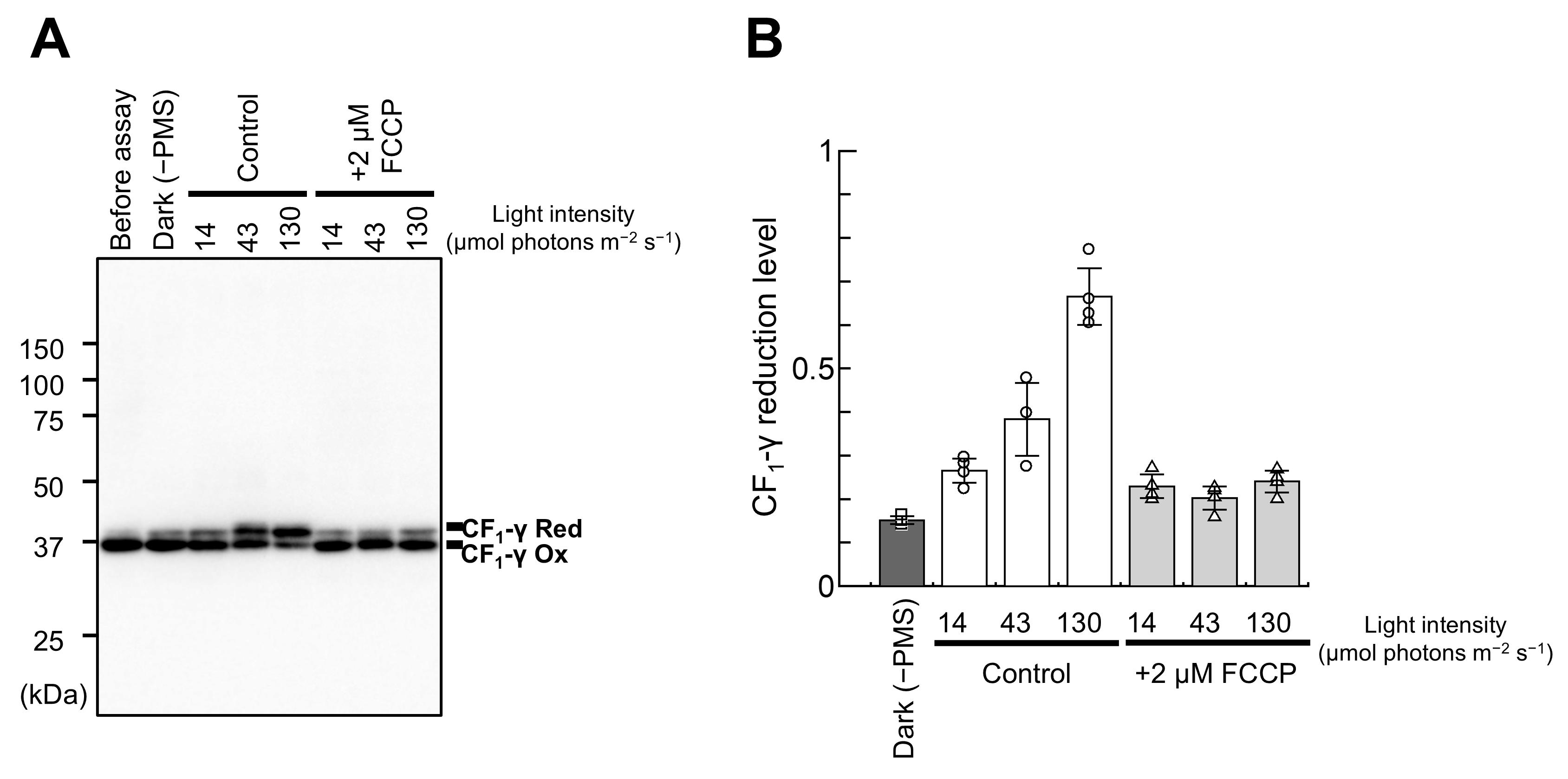
Figure 1. Example results of in vitro CF1-γ reduction using this protocol. (A) Determination of the thylakoid CF1-γ redox state. This protocol reduced CF1-γ in the thylakoids (5 μg Chl/mL) for 5 min. The light intensity of LED irradiation for thylakoids was 14, 43, and 130 μmol photons m-2·s-1. The results were also compared with the case with FCCP added. Ox, oxidized form; Red, reduced form. (B) Quantification of CF1-γ reduction levels for the data shown in (A). Data represent mean ± standard deviation (n = 3–4). Results were quoted with minor modifications from the original paper.
Validation of protocol
This protocol has been used and validated in the following research article:
• Sekiguchi et al. [20]. Proton gradient across the chloroplast thylakoid membrane governs the redox regulatory function of ATP synthase. J. Biol. Chem. 300(9): 107659 (Figure 2).
This protocol is an optimized version of our previous Trx-mediated CFoCF1 reduction protocol, which was used in the following research articles:
• Sekiguchi et al. [18]. Chloroplast ATP synthase is reduced by both f-type and m-type thioredoxins. Biochim. Biophys. Acta Bioenerg. 1861(11):148261 (Figures 1 and 2).
• Sekiguchi et al. [19]. Dissipation of the proton electrochemical gradient in chloroplasts promotes the oxidation of ATP synthase by thioredoxin-like proteins. J. Biol. Chem. 298: 102541 (Figures 2–4).
General notes and troubleshooting
General notes
This protocol provides a robust method for extracting thylakoids from spinach, enzymatic kinetic analysis, and western blotting. Although optimized with specific reagents and conditions, the underlying principles are broadly applicable to plant biochemistry research.
Plant type comparability: The protocol was developed and validated using spinach thylakoids, but its principles are expected to work across other plant types. However, the extraction efficiency of the thylakoids may vary; thus, a minor optimization of the components of the grinding buffer or the strength of homogenizing leaves in the blender may be required.
Detection of Trx target proteins: We successfully detected CF1-γ on the thylakoid membrane using an in-house detection antibody via blotting analysis. If you wish to study a different Trx target protein on the thylakoid membrane, you could simply change the detection antibody.
Troubleshooting
Problem 1: Western blotting showed that CF1-γ appeared to be completely reduced from the beginning of the reaction.
Possible causes: The spinach leaves may not have been kept in the dark long enough before use. Alternatively, the thylakoids may have been exposed to light for too long during isolation.
Solutions: Spinach leaves should always be kept in the dark overnight (or longer) before use, and thylakoid isolation should be performed under dark conditions as much as possible.
Problem 2: During the reaction, the temperature of the reaction solution increases.
Possible cause: The distance between the LED light source used for illumination and the reaction solution may be too short.
Solution: A water bath should be placed between the LED light source and the reaction solution to mitigate an increase in temperature.
Acknowledgments
This protocol was adapted from Sekiguchi et al. [20]. This study was supported by Grants-in-Aid for Scientific Research (Grant 21H02502 to T.H.) from the Japan Society for the Promotion of Science, and partly by a Grant-in-Aid for JSPS Research Fellows (Grant 22J13334 to T.S.) as well as the Dynamic Alliance for Open Innovation Bridging Human, Environment, and Materials.
T.S. and T.H., conceptualization; T.S., investigation; K.Y., resources; T.S., writing—original draft; K.Y. and T.H., writing—review and editing; T.H., supervision.
Competing interests
The authors declare that they have no conflicts of interest regarding the content of this article.
Ethical considerations
All experiments were performed using S. oleracea. No animal or human samples were used.
References
- Junesch, U. and Gräber, P. (1991). The rate of ATP‐synthesis as a function of ΔpH and Δψ catalyzed by the active, reduced H+‐ATPase from chloroplasts. FEBS Lett. 294(3): 275–278. https://doi.org/10.1016/0014-5793(91)81447-g
- Boyer, P. D. (1997). THE ATP SYNTHASE—A SPLENDID MOLECULAR MACHINE. Annu Rev Biochem. 66(1): 717–749. https://doi.org/10.1146/annurev.biochem.66.1.717
- Yoshida, M., Muneyuki, E. and Hisabori, T. (2001). ATP synthase — a marvellous rotary engine of the cell. Nat Rev Mol Cell Biol. 2(9): 669–677. https://doi.org/10.1038/35089509
- Mills, J. D., Mitchell, P. and Schurmann, P. (1980). Modulation of coupling factor ATPase activity in intact chloroplasts, the role of the thioredoxin system. FEBS Lett. 112: 173–177. https://doi.org/10.1016/0014-5793(80)80173-6
- Hisabori, T., Sunamura, E. I., Kim, Y. and Konno, H. (2013). The Chloroplast ATP Synthase Features the Characteristic Redox Regulation Machinery. Antioxid Redox Signal. 19(15): 1846–1854. https://doi.org/10.1089/ars.2012.5044
- Nalin, C. M. and McCarty, R. E. (1984). Role of a disulfide bond in the γ subunit in activation of the ATPase of chloroplast coupling factor 1. J Biol Chem. 259: 7275–7280.
- Miki, J., Maeda, M., Mukohata, Y. and Futai, M. (1988). The γ‐subunit of ATP synthase from spinach chloroplasts Primary structure deduced from the cloned cDNA sequence. FEBS Lett. 232(1): 221–226. https://doi.org/10.1016/0014-5793(88)80421-6
- McKinney, D. W., Buchanan, B. B. and Wolosiuk, R. A. (1978). Activation of chloroplast ATPase by reduced thioredoxin. Phytochemistry. 17(4): 794–795. https://doi.org/10.1016/s0031-9422(00)94230-4
- Mills, J. D. and Mitchell, P. (1982). Modulation of coupling factor ATPase activity in intact chloroplasts. Reversal of thiol modulation in the dark. Biochim Biophys Acta-Bioenerg. 679(1): 75–83. https://doi.org/10.1016/0005-2728(82)90257-2
- Ketcham, S. R., Davenport, J. W., Warncke, K. and McCarty, R. E. (1984). Role of the gamma subunit of chloroplast coupling factor 1 in the light-dependent activation of photophosphorylation and ATPase activity by dithiothreitol. J Biol Chem. 259(11): 7286–7293. https://doi.org/10.1016/s0021-9258(17)39870-8
- Dann, M. S. and McCarty, R. E. (1992). Characterization of the Activation of Membrane-Bound and Soluble CF1 by Thioredoxin. Plant Physiol. 99(1): 153–160. https://doi.org/10.1104/pp.99.1.153
- Jagendorf, A. T. and Uribe, E. (1966). ATP formation caused by acid-base transition of spinach chloroplasts. Proc Natl Acad Sci USA. 55(1): 170–177. https://doi.org/10.1073/pnas.55.1.170
- Junesch, U. and Gräber, P. (1987). Influence of the redox state and the activation of the chloroplast ATP synthase on proton-transport-coupled ATP synthesis/hydrolysis. Biochim Biophys Acta-Bioenerg. 893(2): 275–288. https://doi.org/10.1016/0005-2728(87)90049-1
- Turina, P. (2003). H+/ATP ratio of proton transport-coupled ATP synthesis and hydrolysis catalysed by CF0F1-liposomes. EMBO J. 22(3): 418–426. https://doi.org/10.1093/emboj/cdg073
- Schwarz, O., Schürmann, P. and Strotmann, H. (1997). Kinetics and Thioredoxin Specificity of Thiol Modulation of the Chloroplast H+-ATPase. J Biol Chem. 272(27): 16924–16927. https://doi.org/10.1074/jbc.272.27.16924
- Konno, H., Nakane, T., Yoshida, M., Ueoka-Nakanishi, H., Hara, S. and Hisabori, T. (2012). Thiol Modulation of the Chloroplast ATP Synthase is Dependent on the Energization of Thylakoid Membranes. Plant Cell Physiol. 53(4): 626–634. https://doi.org/10.1093/pcp/pcs018
- Vandermeulen, D. and Govindjee. (1976). Anthroyl stearate as a fluorescent probe of chloroplast membranes. Biochim Biophys Acta-Bioenerg. 449(3): 340–356. https://doi.org/10.1016/0005-2728(76)90146-8
- Sekiguchi, T., Yoshida, K., Okegawa, Y., Motohashi, K., Wakabayashi, K. I. and Hisabori, T. (2020).Chloroplast ATP synthase is reduced by both f-type and m-type thioredoxins. Biochim Biophys Acta-Bioenerg. 1861(11): 148261. https://doi.org/10.1016/j.bbabio.2020.148261
- Sekiguchi, T., Yoshida, K., Wakabayashi, K. I. and Hisabori, T. (2022). Dissipation of the proton electrochemical gradient in chloroplasts promotes the oxidation of ATP synthase by thioredoxin-like proteins. J Biol Chem. 298(11): 102541. https://doi.org/10.1016/j.jbc.2022.102541
- Sekiguchi, T., Yoshida, K., Wakabayashi, K. I. and Hisabori, T. (2024). Proton gradient across the chloroplast thylakoid membrane governs the redox regulatory function of ATP synthase. J Biol Chem. 300(9): 107659. https://doi.org/10.1016/j.jbc.2024.107659
- Porra, R., Thompson, W. and Kriedemann, P. (1989). Determination of accurate extinction coefficients and simultaneous equations for assaying chlorophylls a and b extracted with four different solvents: verification of the concentration of chlorophyll standards by atomic absorption spectroscopy. Biochim Biophys Acta-Bioenerg. 975(3): 384–394. https://doi.org/10.1016/s0005-2728(89)80347-0
- Yoshida, K. and Hisabori, T. (2019). Simple Method to Determine Protein Redox State in Arabidopsis thaliana. Bio Protoc. 9(11): e3250. https://doi.org/10.21769/bioprotoc.3250
Article Information
Publication history
Received: Mar 4, 2026
Accepted: Apr 22, 2026
Available online: May 9, 2026
Published: Jun 5, 2026
Copyright
© 2026 The Author(s); This is an open access article under the CC BY-NC license (https://creativecommons.org/licenses/by-nc/4.0/).
How to cite
Readers should cite both the Bio-protocol article and the original research article where this protocol was used:
- Sekiguchi, T., Yoshida, K. and Hisabori, T. (2026). Evaluating Thioredoxin-Mediated CFoCF1 Reduction Using an In Vitro Thylakoid Assay. Bio-protocol 16(11): e5706. DOI: 10.21769/BioProtoc.5706.
- Sekiguchi, T., Yoshida, K., Wakabayashi, K. I. and Hisabori, T. (2024). Proton gradient across the chloroplast thylakoid membrane governs the redox regulatory function of ATP synthase. J Biol Chem. 300(9): 107659. https://doi.org/10.1016/j.jbc.2024.107659

 Download Citation in RIS Format
Category
Plant Science > Plant biochemistry > Protein > Activity
Biochemistry > Protein > Labeling
Cell Biology > Organelle isolation > Chloroplast
Do you have any questions about this protocol?
Post your question to gather feedback from the community. We will also invite the authors of this article to respond.
 Post a Question


