


- Protocols

- Articles and Issues
- For Authors
- About
- Become a Reviewer


A Feeder Cell-Free System for Chimeric Antigen Receptor Gene Transduction Into Natural Killer Cells

(§Technical contact: nkubo@med.niigata-u.ac.jp) Published: Vol 16, Iss 8, Apr 20, 2026 DOI: 10.21769/BioProtoc.5669 Views: 512
Reviewed by: Alessandro DidonnaAnonymous reviewer(s)
 PDF
PDF  Ask a question
Original research article
The authors used this protocol in:
Oct 2025
Advertisement



Protocol Collections
Comprehensive collections of detailed, peer-reviewed protocols focusing on specific topics




Abstract
Anti-CD19 chimeric antigen receptor (CAR)-natural killer (NK) cells are expected to demonstrate anti-CD19 CAR-T-cell-like efficacy against relapsed and refractory B-cell malignancies and autoimmune diseases, with fewer adverse events and the added advantage of permitting the use of allogeneic cells. However, the methodology for generating CAR-NK cells remains under development. Although various cell sources and expansion methods are available, feeder cells derived from cancerous tissue have been most commonly employed to promote ex vivo expansion of NK cells. In the protocol described herein, NK cells are expanded from adult peripheral blood mononuclear cells using CD2- and NKp46-specific stimulating antibodies in combination with multiple cytokines. The activated NK cells can be genetically modified using a retroviral vector. Subsequent culture of these cells yields large numbers of anti-CD19 CAR-NK cells. The current method, which enables feeder-free, large-scale generation of anti-CD19 CAR-NK cells, eliminates the risk of tumor cell contamination and may facilitate safer clinical application.
Key features
• This method for expanding human primary NK cells ex vivo uses stimulatory antibodies and multiple cytokines, without requiring feeder cells, usually derived from cancerous tissue.
• NK cells are selectively expanded from unsorted peripheral blood mononuclear cells.
• Retroviral vector efficiently mediates gene transfer into NK cells stimulated with the current method.
• Although the cells were not sorted, gene transfer into T cells is minimal.
Keywords: NK cell expansion CAR-NK cells
Anti-NKp46 antibody
Anti-CD2 antibody
Interleukin-12
Interleukin-18
Interleukin-21
Off-the-shelf
CAR-NK cells
Anti-NKp46 antibody
Anti-CD2 antibody
Interleukin-12
Interleukin-18
Interleukin-21
Off-the-shelf
Graphical overview
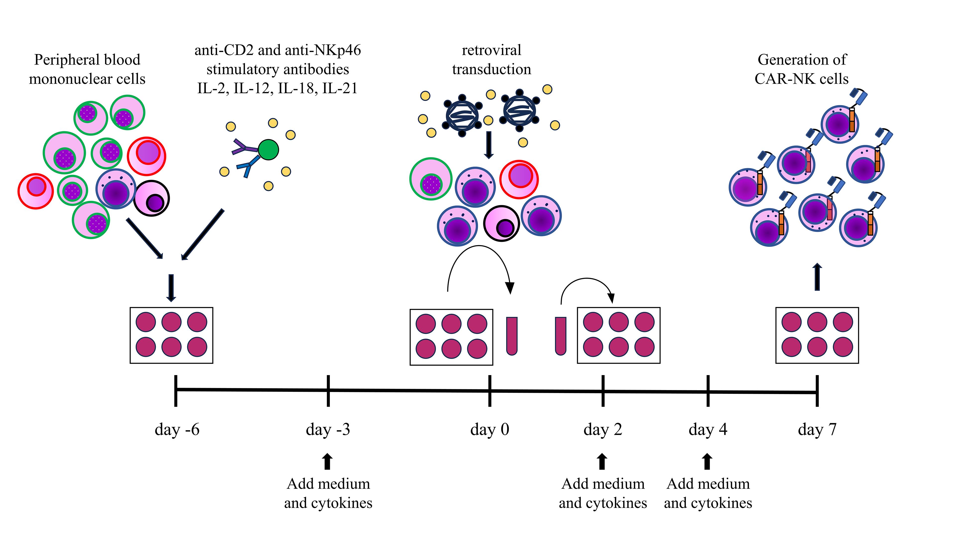
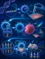
Schematic overview of the generation of chimeric antigen receptor (CAR)-natural killer (NK) cells. Peripheral blood mononuclear cells are collected and stimulated with anti-CD2 and anti-NKp46 stimulatory antibodies in the presence of interleukin (IL)-2, IL-12, IL-18, and IL-21. Cells are cultured in medium supplemented with cytokines, and gene transfer is performed using retroviral vectors after 6 days. The cells are then further cultured under the same conditions to generate CAR-NK cells.
Background
Extensive research continues to establish immune cell therapy as a treatment option for patients with recurrent and/or refractory malignant tumors. This approach has already been realized for certain types of blood cancers and sarcomas. Anti-CD19 chimeric antigen receptor (CAR)-T cells have demonstrated dramatic efficacy against B-cell malignancies [1]. In recent years, anti-CD19 CAR-T cells have also been reported to show promising results in patients with autoimmune diseases, in addition to their use in malignant tumors [2]. Because of allogeneic reactivity, the cell source is typically limited to the patient’s own cells. This approach carries several problems. Insufficient number and poor fitness of T cells collected from heavily treated patients with multiple lines of anti-cancer chemotherapy can result in manufacturing failure [3]. Also, during manufacturing time, which sometimes requires more than a month, progressive disease may result in deterioration of the patient’s performance status and loss of a chance to be infused with CAR-T cells. In addition, cytokine release syndrome (CRS), immune effector cell–associated neurotoxicity syndrome (ICANS), and other immune effector cell–associated complications remain significant concerns [4].
Like anti-CD19 CAR-T cells, anti-CD19 CAR-natural killer (NK) cells are expected to be effective against B-cell malignancies and autoimmune diseases. Furthermore, they offer additional advantages, including the safe use of allogeneic cells and a lower risk of CRS and ICANS [5]. However, the methodology for generating anti-CD19 CAR-NK cells remains insufficiently established. Multiple factors still require optimization, including the choice of cell source, the gene-transduction strategy, and the cell-expansion protocol. Various combinations of cytokines and culture media have been employed to expand NK cells for CAR-NK cell generation [6–8]. Achieving consistent results in expansion efficiency remains challenging. In addition to cytokines, activation with stimulatory antibodies may further enhance expansion efficiency.
We successfully generated anti-CD19 CAR-NK cells from adult peripheral blood mononuclear cells by expanding the cells using antibodies and cytokines, followed by gene transduction via retroviral vectors [9]. Various cell sources have been utilized for CAR-NK cell generation, including umbilical cord blood and induced pluripotent stem (iPS) cells [6,10]. Umbilical cord blood provides a limited volume per collection and contains a relatively small number of NK cells. Although iPS cells have the potential to generate a large number of homogeneous CAR-NK cells, the methodology for their production has not yet been fully established. In contrast, adult peripheral blood mononuclear cells can be collected with relative ease and low cost [11]. Genetically modified tumor cell lines have been used as feeder cells to support ex vivo expansion and genetic modification of NK cells, and this method remains the gold standard for the production of CAR-NK cells [12]. Eliminating the use of feeder cell lines in CAR-NK cell generation removes the risk of tumor cell contamination and enables the large-scale generation of homogeneous CAR-NK cells.
This protocol outlines the procedure for generating anti-CD19 CAR-NK cells from adult peripheral mononuclear cells without the use of feeder cell lines. This protocol can also be applied to the production of various types of engineered NK cells, including CAR-NK cells targeting alternative antigens.
Materials and reagents
Biological materials
1. 293T cell line (American Type Culture Collection, catalog number: CRL-3216)
2. HeLa cell line (American Type Culture Collection, catalog number: CRM-CCL-2)
3. MSCV-CD19 CAR-IRES-GFP plasmids (St. Jude Vector Development and Production Shared Resource [13])
4. pEQ-PAM3 plasmids (St. Jude Vector Development and Production Shared Resource [13])
5. pRDF plasmids (St. Jude Vector Development and Production Shared Resource [13])
6. Human peripheral blood mononuclear cells (collected from healthy adult donors)
Reagents
1. Dulbecco’s modified Eagle’s medium (DMEM), high glucose (Sigma-Aldrich, catalog number: D6429-500ml)
2. Fetal bovine serum (FBS) (Sigma-Aldrich, catalog number: 173012)
3. FuGENE HD transfection reagent (Fugent, catalog number: HD-1000)
4. Roswell Park Memorial Institute (RPMI)-1640 (Fujifilm Wako, catalog number: 189-02025)
5. CellGenix GMP stem cell growth medium (SCGM) (CellGenix Gmb, catalog number: 20802-500)
6. Cloudz Human NK Cell Expansion kit (Bio-Techne, catalog number: CLD004)
7. Recombinant human interleukin (IL)-2 (PeproTech, catalog number:200-02)
8. Recombinant human IL-12 (R&D Systems, catalog number: 10018-IL)
9. Recombinant human IL-18 (R&D Systems, catalog number: 9124-IL)
10. Recombinant human IL-21 (R&D Systems, catalog number: 8879-IL)
11. Polybrene solution (Nacalai Tesque, catalog number: 12996-81)
12. 10× D-PBS (-) (FUJIFILM Wako, catalog number: 048-29805)
13. 30% (w/v) albumin solution, from bovine serum (BSA) (FUJIFILM Wako, catalog number: 017-22231)
14. RetroNectin (Takara, catalog number: T100A)
15. Trypsin–ethylenediaminetetraacetic acid (EDTA) (0.05%), phenol red (Thermo Fisher Scientific Inc., catalog number: 25300-120)
16. Biotin-SP (long spacer) AffiniPure goat anti-mouse IgG, F(ab')2 fragment specific (Jackson ImmunoResearch Laboratories, Inc., catalog number: 115-065-072)
17. Streptavidin-phycoerythrin (BioLegend, catalog number: 405203)
18. Phycoerythrin (PE) anti-human CD56 (NCAM) antibody (BioLegend, catalog number: 318305)
19. Fluorescein isothiocyanate (FITC) anti-human CD3 antibody (BioLegend, catalog number: 300440)
20. Peridinin-chlorophyll-protein complex (PerCP) anti-human CD3 antibody (BioLegend, catalog number: 300427)
21. Acetic acid (FUJIFILM Wako, catalog number: 017-00256)
22. Penicillin-streptomycin solution (100×) (FUJIFILM Wako, catalog number: 168-23191)
23. Ficoll-PaqueTM PLUS (Cytiva, catalog number: 17144002)
24. 0.4% (w/v) Trypan Blue solution (FUJIFILM Wako, catalog number: 207-17081)
25. 4% Paraformaldehyde phosphate buffer solution (FUJIFILM Wako, catalog number: 161-20145)
Solutions
1. DMEM culture medium (see Recipes)
2. RPMI culture medium (see Recipes)
3. SCGM culture medium (see Recipes)
4. 1× PBS (see Recipes)
5. IL-2 solution (see Recipes)
6. IL-12 solution (see Recipes)
7. IL-18 solution (see Recipes)
8. IL-21 solution (see Recipes)
9. 2% BSA (see Recipes)
10. 1× PBS containing 0.5% paraformaldehyde (see Recipes)
Recipes
1. DMEM culture medium
| Reagent | Final concentration | Volume |
|---|---|---|
| DMEM-high glucose | 90% (v/v) | 450 mL |
| FBS (inactivation) | 10% (v/v) | 50 mL |
| Total | n/a | 500 mL |
Before use, FBS is heat-inactivated by incubation at 56 °C for 30 min in a water bath. Store the medium at 4 °C.
2. RPMI culture medium
| Reagent | Final concentration | Volume |
|---|---|---|
| RPMI-1640 | 89% (v/v) | 445 mL |
| FBS (inactivation) | 10% (v/v) | 50 mL |
| Penicillin-streptomycin | 100,000 units/L, 100 mg/L | 5 mL |
| Total | n/a | 500 mL |
Before use, FBS is heat-inactivated by incubation at 56 °C for 30 min in a water bath. Store the medium at 4°C.
3. SCGM culture medium
| Reagent | Final concentration | Volume |
|---|---|---|
| CellGenix GMP SCGM | 90% (v/v) | 450 mL |
| FBS (inactivation) | 10% (v/v) | 50 mL |
| Total | n/a | 500 mL |
Before use, FBS is heat-inactivated by incubation at 56 °C for 30 min in a water bath. Store the medium at 4 °C.
4. 1× PBS
| Reagent | Final concentration | Quantity or volume |
|---|---|---|
| 10× D-PBS (-) | 10% (v/v) | 100 mL |
| Distilled water | 900 mL | |
| Total | n/a | 1,000 mL |
Sterilize the solution by autoclaving at 121 °C for 30 min and store at room temperature.
5. IL-2 solution
| Reagent | Final concentration | Quantity or Volume |
|---|---|---|
| Recombinant human IL-2 | 20 ng/μL | 50 μg |
| Acetic acid | 50 μL | |
| 30% BSA | 82 μL | |
| 1× PBS | 2,368 μL | |
| Total | n/a | 2.5 mL |
Dissolve IL-2 in acetic acid, followed by dilution with PBS containing 0.1% albumin. Store the solution at -20 °C.
6. IL-12 solution
| Reagent | Final concentration | Quantity or volume |
|---|---|---|
| Recombinant human IL-12 | 100 ng/μL | 10 μg |
| 1× PBS | 100 μL | |
| Total | n/a | 100 μL |
Store the solution at -20 °C.
7. IL-18 solution
| Reagent | Final concentration | Quantity or volume |
|---|---|---|
| Recombinant human IL-18 | 200 ng/μL | 10 μg |
| 1× PBS | 50 μL | |
| Total | n/a | 50 μL |
Store the solution at -20 °C.
8. IL-21 solution
| Reagent | Final concentration | Quantity or volume |
|---|---|---|
| Recombinant human IL-21 | 100 ng/μL | 10 μg |
| 1× PBS | 100 μL | |
| 30% BSA | 0.1% (w/v) | 0.3 μL |
| Total | n/a | 100 μL |
Store the solution at -20 °C.
9. 2% BSA
| Reagent | Final concentration | Quantity or volume |
|---|---|---|
| 30% BSA | 2% (w/v) | 1 mL |
| Distilled water | 14 mL | |
| Total | n/a | 15 mL |
Store the solution at 4 °C.
10. 1× PBS containing 0.5% paraformaldehyde
| Reagent | Final concentration | Quantity or volume |
|---|---|---|
| 4% Paraformaldehyde | 0.5% (w/v) | 6 mL |
| 1× PBS | 42 mL | |
| Total | n/a | 48 mL |
Store the solution at 4 °C.
Laboratory supplies
1. NuncTM EasYDISHTM cell culture dishes, tissue culture surface (Thermo Fisher Scientific Inc., catalog number: 150466)
2. Screw cap micro tube, 1.5 mL (SARSTEDT AG & Co., catalog number: 72.692.100)
3. Pipette tip, 0.1–10 μL (Thermo Fisher Scientific Inc, catalog number: 104-Q)
4. Sapphire pipette tip, 1,000 μL (Greiner Bio-One Co., catalog number: 777350)
5. NuncTM biobanking and cell culture tubes, 4.5 mL (Thermo Fisher Scientific Inc., catalog number: 379146)
6. TPP 6-well tissue culture plate (Techno Plastic Products AG, catalog number: 92006)
7. 14 mL round-bottom tube (Corning, catalog number: 352057)
8. 5 mL round-bottom tube (Corning, catalog number: 352052)
9. Parafilm (Bemis, catalog number: PM996)
10. C-Chip disposable hemocytometer (NanoEntek, catalog number: DHC-B02)
11. TPP serological pipette 2 mL (Techno Plastic Products AG, catalog number: 94002)
12. TPP serological pipette 5 mL (Techno Plastic Products AG, catalog number: 94005)
13. TPP serological pipette 10 mL (Techno Plastic Products AG, catalog number: 94010)
14. TPP serological pipette 25 mL (Techno Plastic Products AG, catalog number: 94024)
15. TPP tissue culture flasks 25 (Techno Plastic Products AG, catalog number: 90026)
16. Kimwipe (Crecia, catalog number: 62011)
Equipment
1. Nichipet Air, 1,000 μL (Nichiryo, catalog number: 00-NAR-1000)
2. Nichipet Air, 200 μL (Nichiryo, catalog number: 00-NAR-200)
3. Nichipet Air, 20 μL (Nichiryo, catalog number: 00-NAR-20)
4. Nichipet Air, 10 μL (Nichiryo, catalog number: 00-NAR-10)
5. CO2 incubator (ESPEC, catalog number: BNP-110)
6. Vortex-Genie 2 (IKEDA SCIENTIFIC Co., catalog number: SI-0286)
7. Pipet-Aid XP (Drummond Scientific, catalog number: 4-040-101-J)
8. Hybrid high-speed cooling centrifuge (KUBOTA Corporation Co., catalog number: 6200)
9. BD FACSCalibur flow cytometer (BD Biosciences, catalog number: 342973)
Software and datasets
1. FlowJo v10.0.8 (Becton, Dickson and Company)
Procedure
A. Production of retroviral vector solution
Note: All procedures should be performed under sterile conditions.
(Day -1)
1. Plate 3 × 10⁶ 293T cells into NuncTM EasYDISHTM cell culture dishes containing 10 mL of DMEM culture medium.
2. Incubate the culture dish in a 37 °C and 5% CO2 incubator.
(Day 0)
3. Allow undiluted DMEM high glucose and FuGENE HD transfection reagent to reach room temperature.
4. Add 51 μL of FuGENE HD transfection reagent directly to 800 μL of DMEM in a 1.5 mL screw-cap microcentrifuge tube, taking care to avoid contact with the tube walls. Vortex the tube briefly (approximately 1 s), then incubate it at room temperature for 5 min.
5. Add 6.8 μg of pMSCV-anti-CD19 CAR-IRES-GFP, 6.8 μg of pEQ-PAM3, and 3.4 μg of pRDF to the tube and then vortex the tube briefly (approximately 1 s).
Note: The CAR gene-containing plasmid was constructed based on the MSCV-IRES-GFP plasmid (which can be obtained from Addgene, number: 20672). The anti-CD19 CAR sequence was inserted between the EcoRI and XhoI restriction sites. The pEQ-PAM3 and pRDF plasmids used in this study served as packaging and envelope plasmids, respectively.
6. After incubating at room temperature for 30 min (up to a maximum of 45 min), dispense the mixture in the tube dropwise evenly across the entire surface of the culture medium of 293T cells. Return the culture plate to the incubator and continue incubation.
(Day 1)
7. After 24 h of culture, aspirate and discard the medium from the dishes.
8. Add 10 mL of RPMI culture medium (prewarmed to 37 °C), taking care not to disturb or detach the cell layer.
9. Return the culture plate to the incubator and continue incubation.
(Day 2)
10. As the medium contains the recombinant retroviral particles, collect and aliquot the supernatant of the culture medium into three NuncTM biobanking and cell culture tubes (approximately 3 mL each), and store them at -80 °C. In addition, store a small aliquot (approximately 100 μL) separately at -80 °C for the titration of recombinant retrovirus particles.
11. Immediately after collection, add 10 mL of RPMI culture medium to the dishes, taking care not to disturb or detach the cell layer.
12. Return the culture plate to the incubator and continue incubation.
(Day 3)
13. Again, collect and aliquot the supernatant into three tubes (approximately 3 mL each) and store them at -80 °C. Store a small aliquot (approximately 100 μL) separately at -80 °C for the titration of recombinant retrovirus particles as well.
14. Add 1 mL of Trypsin-EDTA solution to the remaining cells in the dish after washing the cells twice with PBS and incubate the cells in a 37 °C and 5% CO2 incubator for approximately 3 min.
15. Collect the detached 293T cells and disperse them thoroughly by pipetting in RPMI culture medium to reduce cell aggregates and prevent clogging of the fluidics system of the flow cytometry.
Note: Excessive pipetting accelerates cell death; therefore, pipetting should be performed gently and kept to a minimum.
16. After washing the cells with 3 mL of 1× PBS by centrifuging at 190× g for 5 min, resuspend the pelleted cells in 1× PBS containing 0.5% paraformaldehyde, then proceed with flow cytometric analysis. Live cells are gated based on forward scatter (FSC) and side scatter (SSC). The expression of green fluorescent protein (GFP) and CAR protein in the cells indicates the success of transduction and a high probability of generation of recombinant retroviral particles (Figure 1). If only GFP expression is being evaluated in repeated experiments, CAR staining is not required.
Notes:
1. Prepared samples can be stored at 4 °C for up to one week before being analyzed using flow cytometry.
2. The cells, culture media, and washing solutions may contain recombinant retrovirus that do not occur naturally. Therefore, all materials must be autoclaved before disposal.
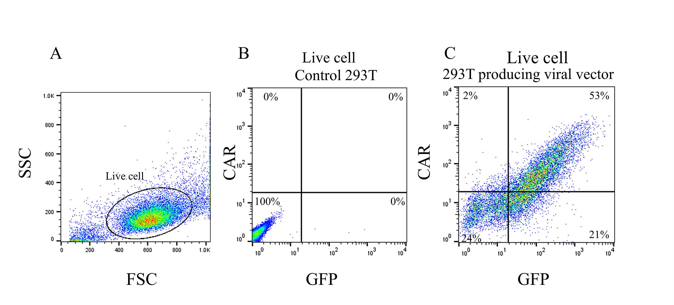
Figure 1. Flow cytometric analysis of viral vector-producing cells. (A) Gated live cells. (B) In control 293T cells, neither green fluorescent protein (GFP) nor chimeric antigen receptor (CAR) was expressed. (C) The 293T cells that successfully produced viral vectors for CAR gene delivery expressed both GFP and CAR.
B. Determination of the titer of recombinant retrovirus particles
Note: All procedures should be performed under sterile conditions.
(Day 0)
1. Plate 1 × 10⁵ HeLa cells in 2 mL of DMEM culture medium per well of a 6-well culture plate and allow to adhere for 4 h.
2. Add any volume (typically 10 to 100 μL) of the stored sample of the culture supernatant to the HeLa cell culture in the presence of polybrene (4 μg/mL). For accurate titration, prepare more than two different conditions (e.g., we typically use 20 and 100 μL in each well), especially when a newly constructed retroviral vector is used.
(Day 5)
3. After washing the cells twice with 1× PBS, add 0.5 mL of Trypsin-EDTA solution and incubate the cells in a 37 °C and 5% CO2 incubator for approximately 3 min.
4. Collect the HeLa cells and disperse them thoroughly by pipetting in RPMI culture medium to reduce cell aggregates and prevent clogging of the fluidics system of the flow cytometry.
5. Wash the cells with 3 mL of 1× PBS by centrifuging at 190× g for 5 min and resuspend the cell pellet in 1× PBS containing 0.5% paraformaldehyde. Then, proceed with flow cytometric analysis. As only GFP expression is being evaluated, staining is unnecessary.
6. Determine the percentage of GFP-positive cells in the live cell gate (FSC/SSC). The titer is calculated using the following formula:
Recombinant retrovirus titer (particles in 1 mL of the conditioned medium) = A/100 × 1,000/B × 105
where A is the percentage of GFP-positive cells (%), and B is the volume of the aliquot (μL) (Figure 2). If different values are obtained from independent wells exposed to different volumes of the retroviral supernatants, the higher one may be appropriate. According to our personal experience, the percentage of GFP-positive HeLa cells usually ranges from 1% to 15% in ideal conditions. In cases involving over 15% GFP-positive cells, it is possible that more than one copy of the viral genome has integrated into a single cell, where an underestimation of the titer of the recombinant retroviral particles may occur.
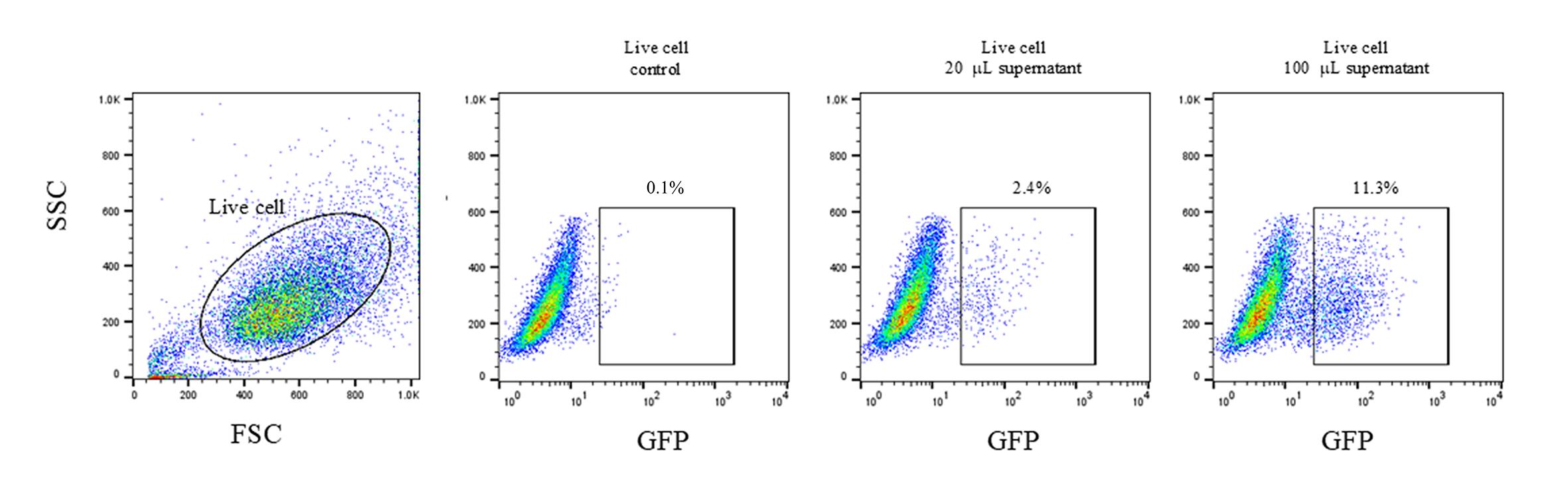
Figure 2. Determination of viral titer by flow cytometry. Live cells were gated based on forward (FSC) and side scatter (SSC). The percentage of green fluorescent protein (GFP)-positive cells was determined among the gated live cell population. The proportion of GFP-positive cells increased as the volume of retroviral supernatants increased. The respective titer calculations are 1.2 × 10⁵ particles/mL for GFP+ 2.4% and 1.13 × 10⁵ particles/mL for GFP+ 11.3%.
C. Generation of anti-CD19 CAR-NK cells
Note: All procedures should be performed under sterile conditions.
(Day -6)
1. Isolate peripheral blood mononuclear cells (PBMCs) from heparinized blood obtained from healthy adult donors using Ficoll density gradient centrifugation, according to the protocol described by Panda et al. [14].
2. Wash the PBMCs with 5 mL of RPMI-1640 with no additives by centrifuging at 190× g for 5 min. Resuspend the cells in SCGM culture medium prewarmed to 37 °C. Count the cells using the Trypan Blue dye exclusion method.
3. Transfer a small portion (e.g., 2–5 × 10⁴) of the isolated PBMCs into a 5 mL round-bottom tube. Add fluorescently labeled antibodies against CD3 and CD56 according to the manufacturer’s instructions and incubate as directed. Wash the cells with 1 × PBS by centrifuging at 190× g for 5 min and resuspend the pelleted cells in PBS. Determine the proportion of NK cells and CD3-negative and CD56-positive lymphocytes within live cell gating by flow cytometry.
4. Prepare 4 mL of SCGM culture medium prewarmed to 37 °C containing 4 × 10⁵ NK cells.
5. Add 5.4 μL of IL-2 solution (final concentration of 27 ng/mL), 0.4 μL of IL-12 solution (final concentration of 10 ng/mL), 0.2 μL of IL-18 solution (final concentration of 10 ng/mL), and 0.4 μL of IL-21 solution (final concentration of 10 ng/mL) to 4 mL of the cell suspension.
6. Vortex the entire vial of Cloudz CD2/NKp46 for 30 s. Add 4.5 μL of the reagent to 4 mL of the cell suspension. Mix thoroughly by pipetting up and down five times.
7. Add 2 mL of the cell suspension to each of two wells in a 6-well tissue culture plate and place the culture plate in an incubator.
Note: The culture can also be initiated by adjusting the cell number and the medium volume in a T25 flask. For example, a culture may be started with 6 × 10⁴ NK cells in 8 mL of medium by placing the flask vertically.
(Day -3)
8. Prepare 2 mL of SCGM culture medium containing 60 ng/mL IL-2, 30 ng/mL IL-12, 30 ng/mL IL-18, and 30 ng/mL IL-21.
9. Add 1 mL of this mixture to each well (in two wells) to achieve the following final concentrations: 20 ng/mL IL-2, 10 ng/mL IL-12, 10 ng/mL IL-18, and 10 ng/mL IL-21.
Note: The volume of medium may be adjusted according to the cell proliferation.
(Day 0)
10. To prepare a RetroNectin-coated tube, mix 50 μL of RetroNectin (1 mg/mL, pre-dissolved at room temperature) and 450 μL of 1 × PBS to obtain a total volume of 500 μL (100 μg/mL) in a 14 mL round-bottom tube.
11. Incubate the tube at room temperature for 2 h.
Note: Alternatively, the tube may be incubated at 4 °C overnight.
12. Remove the RetroNectin solution (and transfer the solution to a new 14 mL round-bottom tube if needed) and then add 500 μL of 2% BSA in 1× PBS to the RetroNectin-coated tube. Incubate the tube at room temperature for 30 min for blocking of nonspecific binding.
Note: The diluted RetroNectin solution (transferred to a new tube) can be used to prepare multiple RetroNectin-coated tubes by continuing from step C11.
13. Discard the blocking solution, wash the tube once with 2 mL of 1× PBS, and use it for retroviral transduction.
Note: After removing the buffer, seal the RetroNectin-coated tube tightly with Parafilm. It can be stored at 4 °C for up to one week. Accordingly, steps C10–13 may be completed in advance.
14. Add appropriate volume (e.g., 1 mL) of conditioned medium containing recombinant retrovirus particles prepared in section A to the RetroNectin-coated tube and centrifuge at 760× g for 120 min at 32 °C. Discard the viral supernatant and wash the tube once with 2 mL of 1 × PBS.
15. Collect the mononuclear cells that have been cultured for 6 days and centrifuge them at 190× g for 5 min at 20 °C. Discard the supernatant and resuspend the cells in 1 mL of SCGM culture medium supplemented with 20 ng/mL IL-2, 10 ng/mL IL-12, 10 ng/mL IL-18, and 10 ng/mL IL-21. Transfer the cells to a RetroNectin-coated tube and incubate in a 37 °C and 5% CO2 incubator.
Note: Ideally, adjust the viral inoculum volume and NK cell count (approximately 1–2 × 10⁵) to achieve at least a multiplicity of infection (MOI) of 2.
(Day 2)
16. Transfer the cells and culture medium from the RetroNectin-coated tube to a well of a 6-well tissue culture plate.
17. Prepare 2 mL of SCGM culture medium containing 40 ng/mL IL-2, 20 ng/mL IL-12, 20 ng/mL IL-18, and 20 ng/mL IL-21.
18. Add 1 mL of this mixture to each well to achieve the following final concentrations: 20 ng/mL IL-2, 10 ng/mL IL-12, 10 ng/mL IL-18, and 10 ng/mL IL-21.
Note: The volume of medium may be adjusted according to cell proliferation.
(Day 4)
19. Prepare 4 mL of SCGM culture medium containing 40 ng/mL IL-2, 20 ng/mL IL-12, 20 ng/mL IL-18, and 20 ng/mL IL-21.
20. Add 2 mL of this mixture to each well to achieve the following final concentrations: 20 ng/mL IL-2, 10 ng/mL IL-12, 10 ng/mL IL-18, and 10 ng/mL IL-21.
Note: The volume of medium may be adjusted according to the cell proliferation.
(Day 7)
21. Collect the cultured cells and assess cell count and transduction efficiency (GFP positivity and CAR expression) by flow cytometry.
22. For continued culture, replenish the medium every 2–3 days by adding the IL-2 stock solution to achieve a final concentration of 20 ng/mL.
Data analysis
Generated anti-CD19 CAR-NK cells are analyzed by flow cytometry. Collect a small aliquot (approximately 100 μL) in a 5 mL round-bottom tube, wash the cells with 3 mL of 1× PBS by centrifuging at 190× g for 5 min, and resuspend the pelleted cells in 1× PBS. Add the fluorescently labeled antibodies to the tube according to the manufacturer’s instructions. After incubation as directed, wash the cells with 3 mL of 1× PBS by centrifuging at 190× g for 5 min. Resuspend the pelleted cells in 1× PBS (or 1× PBS containing 0.5% paraformaldehyde if analysis is not immediate) and proceed with flow cytometric analysis. Lymphocytes are gated based on FSC and SSC to assess GFP expression and the fluorescence of each marker. Because GFP is expressed in transduced cells, FITC-conjugated antibodies cannot be used. Alternative fluorochromes such as PE or PerCP may be used instead. To assess NK cell expansion and confirm the absence of T-cell contamination, use antibodies against CD56 and CD3. CAR expression is assessed using antibodies targeting the extracellular domain (mouse immunoglobulin G) or a fluorochrome-conjugated target protein (e.g., CD19 conjugated with PE). For example, CAR expression can be detected using Biotin-SP AffiniPure goat anti-mouse IgG, followed by streptavidin-phycoerythrin, irrespective of the CAR specificity (Figure 3).
Note: Prepared samples can be stored at 4 °C for up to one week and analyzed using flow cytometry at a later date.
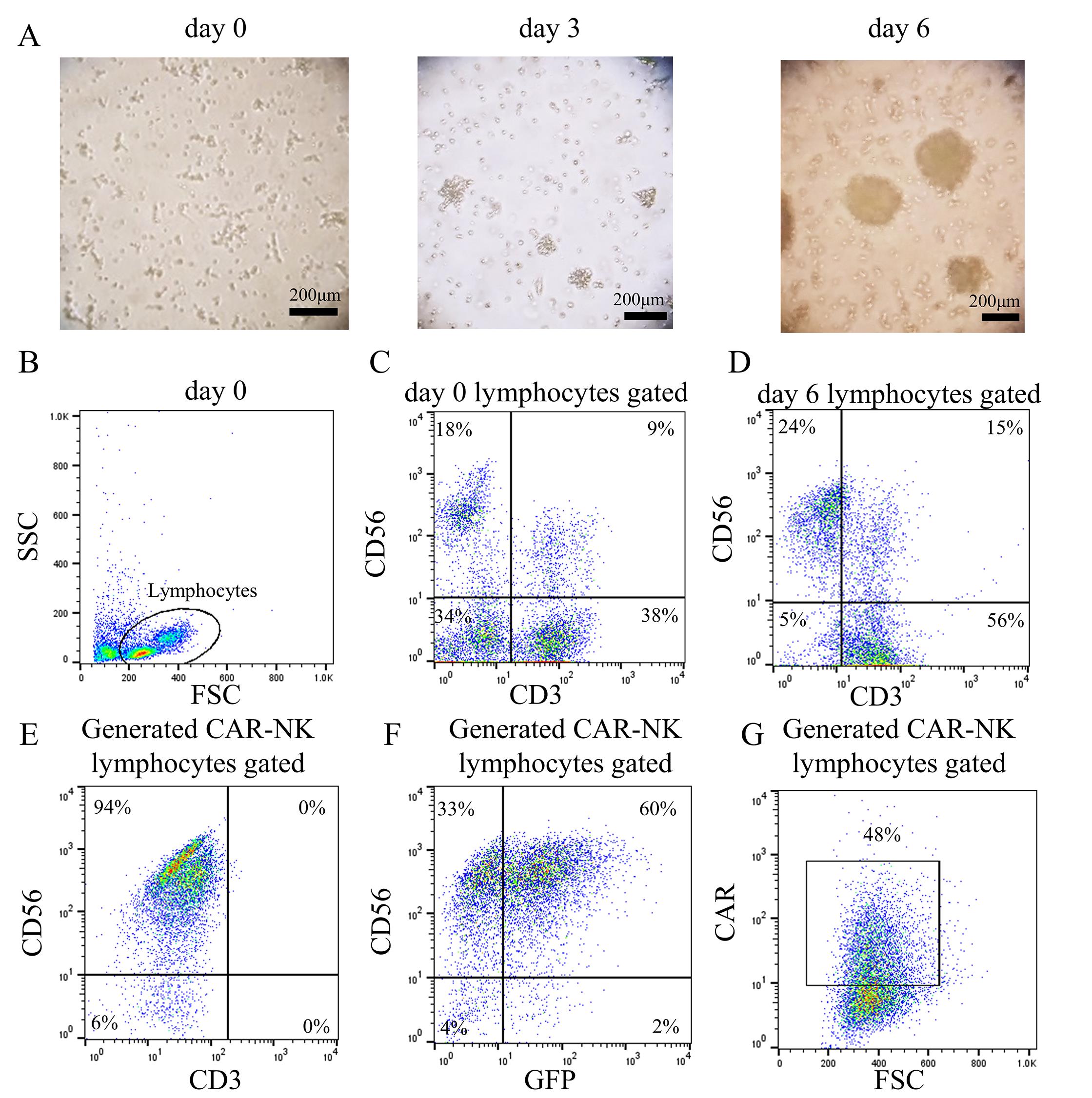
Figure 3. Successful generation of chimeric antigen receptor (CAR)-natural killer (NK) cells. (A) Microscopic image of cells during culture. Scale bar: 200 μm. (B–G) Flow cytometric analysis before culture, during culture, and after generation of CAR-NK cells. (B) Gating strategy of lymphocytes. Side (SSC) and forward scatter (FSC) are shown on the y-axis and x-axis, respectively. (C) CD56 and CD3 expression prior to culture are shown on the y-axis and x-axis, respectively. (D) CD56 and CD3 expression during culture are shown on the y-axis and x-axis, respectively. Note that NK cell expansion is not significant on day 6 of the culture process. However, massive expansion is expected on day 10 or later (see [9]). (E) CD56 and CD3 expression of CAR-NK cells are shown on the y-axis and x-axis, respectively. (F) CD56 and green fluorescent protein (GFP) expression of CAR-NK cells are shown on the y-axis and x-axis, respectively. (G) CAR expression analyzed using antibodies against the extracellular domain of mouse immunoglobulin.
Validation of protocol
This protocol has been used and validated in the following research article:
Kubo et al. [9]. A novel method for generating chimeric antigen receptor-transduced primary human natural killer cells by the use of multiple cytokines and anti-CD2 and anti-NKp46 stimulatory antibodies. Biomed Pharmacother. 191: 118505. doi: 10.1016/j.biopha.2025.118505.
In the original paper, we investigated the optimal timing for gene transfer following the initiation of NK cell expansion culture using unsorted peripheral blood mononuclear cells. The timing outlined in this protocol yielded the most efficient transfer. Gene transfer of the anti-CD19 CAR construct was performed using the protocol described herein. Transduction efficiency was 57.9% ± 14.6% (mean ± standard deviation; n = 6) on day 7 post-transduction. The number of GFP-positive NK cells increased by 16.6 ± 8.6-fold after 7 days.
General notes and troubleshooting
General notes
1. Interindividual variation in NK cell expansion efficiency: NK cell expansion efficiency using this protocol varies markedly among individuals. Individuals with low expansion capacity consistently show poor outcomes, even when the same protocol is repeated. The extent of NK cell expansion cannot be predicted in advance for a given individual. Therefore, it is advisable to assess NK cell expansion for each individual prior to gene transfer.
2. Genes that can be transduced via this protocol are not limited to the anti-CD19 CAR construct: This protocol can be utilized to introduce various genes into NK cells. It is applicable to CAR constructs targeting antigens other than CD19, as well as genes encoding membrane-bound cytokines.
3. Because human-derived samples may be contaminated with infectious organisms, they should be handled in accordance with institutional biosafety guidelines. Adherence to appropriate biosafety levels and wearing of personal protective equipment is essential.
4. Based on our experience, we know that recombinant retroviral supernatants stored at -80 °C for more than one year retain their ability to transduce efficiently.
5. The 293T cell line should not be passaged beyond approximately 10 passages.
Troubleshooting
Problem 1: Low transduction efficiency in NK cells.
Possible cause: Low retrovirus titer.
Solution: Overconfluent 293T cells readily form cell clumps, which reduces transfection efficiency during virus production. To prevent this, cells should be passaged with trypsin before reaching 80% confluence.
Problem 2: Low yield of anti-CD19 CAR-NK cells.
Possible cause: Interindividual variability in NK cell expansion efficiency.
Solution: Select peripheral blood donors who have previously shown good results.
Acknowledgments
Conceptualization, C.I.; Methodology, Y.K., R.H.; Investigation, N.K., M.B., Y.S.; Writing—Original Draft, N.K.; Writing—Review & Editing, C.I.; Funding acquisition, N.K., M.I., C.I.; Supervision, A.S.
This study was supported by the Japan Society for the Promotion of Science (JSPS) Grants-in-Aid for Scientific Research, JSPS KAKENHI [to C.I. (19K08137, 22H03036, 25K02622) and to M.I. (21K07213)], the Morinaga Foundation for Health & Nutrition Research Fund to N.K., and the Japanese Society of Hematology Research Grant to C.I.
This protocol was initially described in Kubo et al. [9]. The authors would like to acknowledge the use of ChatGPT 5.2 for the purpose of language editing and grammatical correction to improve the readability of the manuscript. Generative AI was not used in the conceptualization or analytical processes of this study.
Competing interests
Chihaya Imai reports a relationship with CURED Inc. that includes consulting or advisory and funding grants. Chihaya Imai reports patent royalties from Juno Therapeutics. Yuko Suzuki is a former employee of CURED, Inc. The other authors declare that they have no known competing financial interests or conflicts of interest.
Ethical considerations
Written informed consent was obtained from all healthy adult volunteers who donated peripheral blood samples. The study was conducted in accordance with the guidelines of the Declaration of Helsinki and was approved by the ethics committee of Niigata University School of Medicine (approval #2015-2686).
References
- Maude, S. L., Laetsch, T. W., Buechner, J., Rives, S., Boyer, M., Bittencourt, H., Bader, P., Verneris, M. R., Stefanski, H. E., Myers, G. D., et al. (2018). Tisagenlecleucel in Children and Young Adults with B-Cell Lymphoblastic Leukemia. N Engl J Med. 378(5): 439–448. https://doi.org/10.1056/nejmoa1709866
- Müller, F., Taubmann, J., Bucci, L., Wilhelm, A., Bergmann, C., Völkl, S., Aigner, M., Rothe, T., Minopoulou, I., Tur, C., et al. (2024). CD19 CAR T-Cell Therapy in Autoimmune Disease — A Case Series with Follow-up. N Engl J Med. 390(8): 687–700. https://doi.org/10.1056/nejmoa2308917
- Baguet, C., Larghero, J. and Mebarki, M. (2024). Early predictive factors of failure in autologous CAR T-cell manufacturing and/or efficacy in hematologic malignancies. Blood Adv. 8(2): 337–342. https://doi.org/10.1182/bloodadvances.2023011992
- Lee, D. W., Santomasso, B. D., Locke, F. L., Ghobadi, A., Turtle, C. J., Brudno, J. N., Maus, M. V., Park, J. H., Mead, E., Pavletic, S., et al. (2019). ASTCT Consensus Grading for Cytokine Release Syndrome and Neurologic Toxicity Associated with Immune Effector Cells. Biol Blood Marrow Transplant. 25(4): 625–638. https://doi.org/10.1016/j.bbmt.2018.12.758
- Marin, D., Li, Y., Basar, R., Rafei, H., Daher, M., Dou, J., Mohanty, V., Dede, M., Nieto, Y., Uprety, N., et al. (2024). Safety, efficacy and determinants of response of allogeneic CD19-specific CAR-NK cells in CD19+ B cell tumors: a phase 1/2 trial. Nat Med. 30(3): 772–784. https://doi.org/10.1038/s41591-023-02785-8
- Herrera, L., Santos, S., Vesga, M. A., Anguita, J., Martin-Ruiz, I., Carrascosa, T., Juan, M. and Eguizabal, C. (2019). Adult peripheral blood and umbilical cord blood NK cells are good sources for effective CAR therapy against CD19 positive leukemic cells. Sci Rep. 9(1): 18729. https://doi.org/10.1038/s41598-019-55239-y
- Oberschmidt, O., Morgan, M., Huppert, V., Kessler, J., Gardlowski, T., Matthies, N., Aleksandrova, K., Arseniev, L., Schambach, A., Koehl, U., et al. (2019). Development of Automated Separation, Expansion, and Quality Control Protocols for Clinical-Scale Manufacturing of Primary Human NK Cells and Alpharetroviral Chimeric Antigen Receptor Engineering. Hum Gene Ther Methods. 30(3): 102–120. https://doi.org/10.1089/hgtb.2019.039
- Müller, S., Bexte, T., Gebel, V., Kalensee, F., Stolzenberg, E., Hartmann, J., Koehl, U., Schambach, A., Wels, W. S., Modlich, U., et al. (2020). High Cytotoxic Efficiency of Lentivirally and Alpharetrovirally Engineered CD19-Specific Chimeric Antigen Receptor Natural Killer Cells Against Acute Lymphoblastic Leukemia. Front Immunol. 10: e03123. https://doi.org/10.3389/fimmu.2019.03123
- Kubo, N., Baba, M., Shin, C., Kasahara, Y., Hosokai, R., Murayama, Y., Oike, N., Mihara, K., Saitoh, A., Imamura, M., et al. (2025). A novel method for generating chimeric antigen receptor-transduced primary human natural killer cells by the use of multiple cytokines and anti-CD2 and anti-NKp46 stimulatory antibodies. Biomed Pharmacother. 191: 118505. https://doi.org/10.1016/j.biopha.2025.118505
- Lin, X., Guan, T., Xu, Y., Li, Y., Lin, Y., Chen, S., Chen, Y., Wei, X., Li, D., Cui, Y., et al. (2024). Efficacy of the induced pluripotent stem cell derived and engineered CD276-targeted CAR-NK cells against human esophageal squamous cell carcinoma. Front Immunol. 15: e1337489. https://doi.org/10.3389/fimmu.2024.1337489
- Wang, F., Huang, Z., Feng, X. and Shi, M. (2025). Immunotherapy of chimeric antigen receptor NK cells: status and its promising future. Front Immunol. 16: e1608277. https://doi.org/10.3389/fimmu.2025.1608277
- Imai, C., Iwamoto, S. and Campana, D. (2005). Genetic modification of primary natural killer cells overcomes inhibitory signals and induces specific killing of leukemic cells. Blood. 106(1): 376–383. https://doi.org/10.1182/blood-2004-12-4797
- Imai, C., Mihara, K., Andreansky, M., Nicholson, I. C., Pui, C. H., Geiger, T. L. and Campana, D. (2004). Chimeric receptors with 4-1BB signaling capacity provoke potent cytotoxicity against acute lymphoblastic leukemia. Leukemia. 18(4): 676–684. https://doi.org/10.1038/sj.leu.2403302
- Panda, S. and Ravindran, B. (2013). Isolation of Human PBMCs. Bio Protoc. 3(3): e323. https://doi.org/10.21769/bioprotoc.323
Article Information
Publication history
Received: Jan 22, 2026
Accepted: Mar 17, 2026
Available online: Apr 1, 2026
Published: Apr 20, 2026
Copyright
© 2026 The Author(s); This is an open access article under the CC BY-NC license (https://creativecommons.org/licenses/by-nc/4.0/).
How to cite
Kubo, N., Baba, M., Suzuki, Y., Kasahara, Y., Hosokai, R., Imamura, M., Saitoh, A. and Imai, C. (2026). A Feeder Cell-Free System for Chimeric Antigen Receptor Gene Transduction Into Natural Killer Cells. Bio-protocol 16(8): e5669. DOI: 10.21769/BioProtoc.5669.

 Download Citation in RIS Format
Category
Immunology > Immunotherapy
Cancer Biology > Tumor immunology > Cancer therapy
Immunology > Immune cell isolation > Maintenance and differentiation
Do you have any questions about this protocol?
Post your question to gather feedback from the community. We will also invite the authors of this article to respond.
 Post a Question


