


- Protocols

- Articles and Issues
- For Authors
- About
- Become a Reviewer


Uptake Assay of Ram Seminal Plasma Extracellular Vesicles to Sperm

Published: Vol 16, Iss 7, Apr 5, 2026 DOI: 10.21769/BioProtoc.5653 Views: 408
Reviewed by: Noelia ForesiMarisa Conte
 PDF
PDF  Ask a question
Original research article
The authors used this protocol in:

Feb 2026
Advertisement



Protocol Collections
Comprehensive collections of detailed, peer-reviewed protocols focusing on specific topics




Related protocols
Abstract
Extracellular vesicles (EVs) are critical mediators of cell–cell communication and play a key role in male reproductive biology by modulating sperm function. This protocol describes a robust and reproducible workflow for isolating EVs from ram seminal plasma using size-exclusion chromatography (SEC) and assessing their uptake by ram spermatozoa. In contrast to ultracentrifugation-based methods, SEC provides a gentle and more efficient isolation approach that preserves EV integrity and functionality. A central innovation of this protocol is the use of carboxyfluorescein succinimidyl ester (CFSE)-labeled seminal plasma EVs (SP-EVs) to evaluate their incorporation into sperm cells through two complementary detection platforms: (i) flow cytometry with standard resolution and (ii) confocal microscopy, for spatial confirmation of EV–sperm interactions. By bridging the gap between EV isolation and functional analysis, this protocol provides a valuable tool for investigating the role of EV–cell interactions. Specifically, it offers potential applications in male fertility preservation, biomarker discovery, and the development of EV-based therapeutic strategies in reproductive medicine.
Key features
• Provides a gentle, SEC-based EV isolation method optimized for ram seminal plasma, suitable for preserving vesicle integrity in studies of male reproductive biology.
• Integrates EV purification with functional assays, enabling direct evaluation of EV–sperm interactions through confocal microscopy and flow cytometry.
• Includes a reproducible CFSE-labeling strategy tailored for seminal plasma EVs, ensuring consistent detection of vesicle uptake by ram spermatozoa.
• Designed for applications in fertility research, offering a workflow compatible with biomarker discovery, cryopreservation studies, and development of EV-based reproductive interventions.
Keywords: Extracellular vesicles Seminal plasma
Sperm
Size-exclusion chromatography
CFSE
Flow cytometry
Seminal plasma
Sperm
Size-exclusion chromatography
CFSE
Flow cytometry
Graphical overview
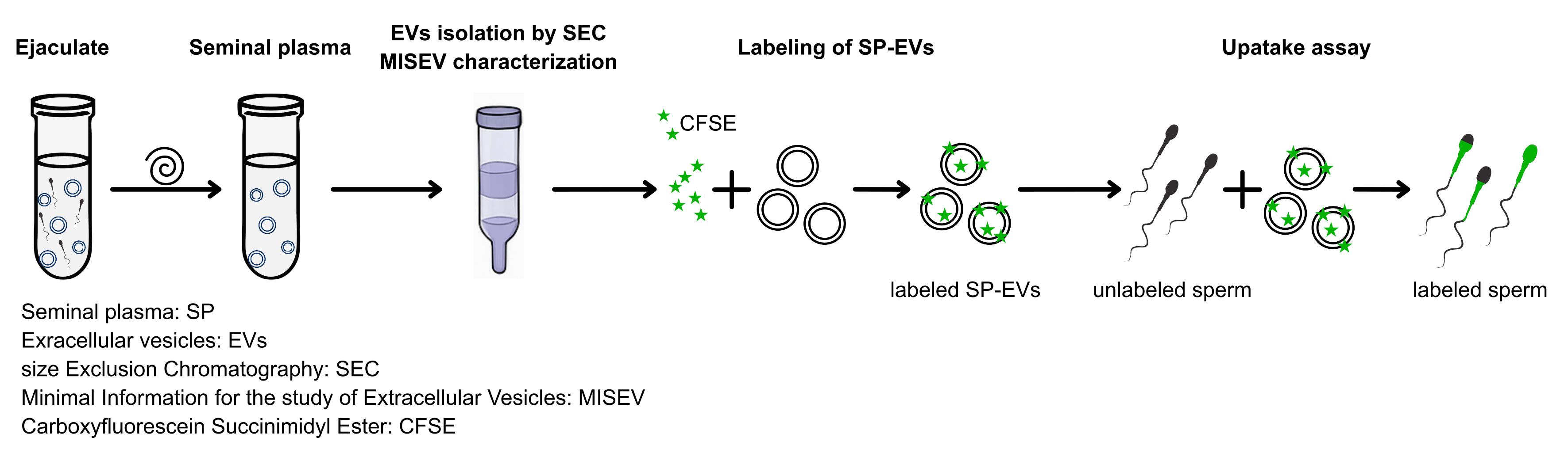
Extracellular vesicles uptake assay by CFSE labeling
Background
Extracellular vesicles (EVs) are membrane-bound particles present in all biological fluids that act as key mediators of intercellular communication [1]. By transporting proteins, lipids, and nucleic acids, they regulate a wide range of cellular processes [1–3]. In reproductive biology, EVs derived from seminal plasma have gained prominence as pivotal regulators of sperm function, exerting influence on processes including motility, capacitation, and fertilization [4–8]. Additionally, EVs from seminal plasma have also been demonstrated to have a subsequent impact on preimplantation, implantation, post-implantation, and pregnancy-associated diseases, as well as on seminal infections [9]. However, despite their relevance, progress in this field has been limited due to methodological variability and lack of standardized, reproducible assays for assessing EV–sperm interactions at the molecular level [10,11].
To address this gap, the present protocol provides an integrated workflow that goes beyond EV isolation and introduces a robust assay to evaluate the uptake of carboxyfluorescein succinimidyl ester (CFSE)-labeled seminal plasma EVs by ram spermatozoa. The key innovation lies in the labeling of seminal plasma (SP)-EVs with CFSE, their co-incubation with sperm cells, and the subsequent detection of vesicle internalization. While EV identity is validated through standard characterization methods—transmission electron microscopy (TEM) and nanoparticle tracking analysis (NTA)—all subsequent steps of the uptake assay can be performed in a conventional laboratory equipped with basic flow cytometry and fluorescence microscopy.
The potential applications of this protocol are manifold for fundamental research on EV–cell interactions in male infertility. This protocol aims to advance the field by providing a reliable tool for studying EVs in reproductive biology and beyond.
Materials and reagents
Biological materials
1. Ram semen samples
2. Semen straws
Reagents
1. Phosphate-buffered saline (PBS) (Gibco, catalog number: 10010049)
2. Primary antibodies: anti-CD9 (1:1000, Sigma-Aldrich, catalog number: A4Y16), anti-CD63 (1:2,000, Cell Signal, catalog number: 55051), anti-HSP70 (1:1,000, Sigma-Aldrich, catalog number: SAB420071), anti-CNX (1:1,000, Abcam, catalog number: ab22595), and anti-ApoC3 (1:3,000, Invitrogen, catalog number: PA5-114865)
3. Secondary antibodies: anti-mouse IgG-HRP conjugated (1:10,000, Sigma-Aldrich, catalog number: A4Y16) and anti-rabbit IgG-HRP conjugated (1:10,000, Sigma-Aldrich, catalog number: A615Y)
4. Dextran blue solution (1 mg/mL in PBS) (Sigma-Aldrich, catalog number: D5751)
5. Sperm selection density gradient (e.g., Ovipure, Nidacon Laboratories AB, Göthenborg, Sweden; or Percoll, Sigma-Aldrich, catalog number: P4937)
6. CellTrace CFSE Cell Proliferation kit (Thermo Fisher Scientific, catalog number: C34554 A); contains dimethyl sulfoxide (DMSO)
7. 10% and 15% SDS-PAGE pre-cast gels (Bio-Rad, catalog number: 456-8033)
8. Ethanol (Sigma-Aldrich, catalog number: 51976)
Laboratory supplies
1. Pipettes (any brand)
2. Tubes (Eppendorf, catalog number: 0030120086)
3. Ultracentrifuge tubes (Beckman Coulter, catalog number: 355631)
4. SuperdexTM 200 column (Cytiva, formerly Amersham Pharmacia, catalog number: 17-1043-01)
5. 0.22 μm filters (Millipore, catalog number: SLGP033RS)
6. PVDF membrane (Millipore, catalog number: IPVH00010)
7. Neubauer chamber (Boeco, Germany)
8. Microscope slides (Thermo Fisher, catalog number: 9951B9)
Equipment
1. Centrifuge (Eppendorf, model: 5810 R)
2. Ultracentrifuge (Beckman Coulter, model: Optima LE-80k)
3. Rotor type 90 Ti (Beckman Coulter, catalog number: 355530)
4. Spectrophotometer (Nanodrop One Thermo Fisher Scientific, catalog number: ND-ONE-W)
5. Flow cytometer (any brand)
6. Confocal microscope (any brand)
7. Water bath (any brand)
8. Heating block (any brand)
9. Rotating mixer (any brand)
10. pH meter (any brand)
11. Gel electrophoresis vertical apparatus (any brand)
12. Semi-dry protein gel transfer system (for gels up to 9 × 6 cm, any brand/model)
13. Chemiluminescence scanner (LI-COR)
14. SEC column stand/rack
15. Peristaltic pump (any brand) (optional)
Software and datasets
1. Licor C-Digit chemiluminescence scanner version 5.2.0
2. GraphPad Prism version 8.0.2
3. RStudio version 3.3.3
4. ImageJ (https://imagej.nih.gov/ij/)
5. Flow cytometry software
Procedure
A. Seminal plasma preparation
1. Collect ejaculates using an artificial vagina. Pool samples according to sperm mass motility (≥4; scale 1–5).
2. Centrifuge the semen sample at 800× g for 10 min at 4 °C. Collect the supernatant, avoiding disturbing the sperm pellet as much as possible, to remove the majority of spermatozoa.
3. Centrifuge the supernatant twice at 16,000× g for 30 min at 4 °C. Collect the supernatant, being careful not to disturb the pellet, to remove cells and cellular debris, ensuring minimal contamination from the pellet.
4. Examine 5 μL of the supernatant under a microscope to confirm the absence of cells and debris.
5. Seminal plasma can be used fresh or aliquoted into desired volumes and stored at -80 °C. If the plasma is to be used within 7 days, it can be stored at 4 °C.
B. Isolation of EVs by size-exclusion chromatography (SEC)
1. Column preparation (Figure 1): Assemble the SuperdexTM 200 column (10 mm inner diameter × 10 cm length; total bed volume 10 mL) and ensure it is homogeneous. During assembly and setting, keep the column wet by adding 0.22 μm filtered-PBS (pH 7.4) to prevent resin from drying.
Note: To preserve column performance and resin integrity, ensure that the column remains fully hydrated at all times. After each run, wash with 2 column volumes of PBS to remove residual samples. For longer storage (>2 days), flush the column with 2 volumes of 20% ethanol in distilled water as a storage solution. Next, seal the column with end caps or connect tubing between the inlet and outlet to prevent air entry and resin drying, and store the column at 4 °C. Do not store the column in NaOH or other harsh cleaning solutions. Before reuse, equilibrate the column with at least 2 volumes of PBS.
2. Equilibrate the column with 1–2 volumes of filtered PBS at 4 °C. Use PBS as a running buffer. Note the manufacturer’s recommended flow range and do not exceed it. If not specified, use 1–3 mL/min flow rate.
3. For void volume (V0) determination, use 400 μL of dextran blue solution (1 mg/mL in PBS). Before adding dextran blue, make sure to remove any excess liquid from the top of the column. After the dextran has entered the bed, immediately top up with PBS to avoid any air entry or drying.
4. Collect 0.5 mL fractions and record the elution profile of dextran blue. Calculate the void volume as the fraction(s) where the dextran blue elutes.
5. Equilibrate the column again by passing 1–2 volumes of filtered PBS.
6. For sample application, remove any excess liquid from the top of the column and apply 400 μL of seminal plasma. Allow the plasma to enter the column completely before adding PBS.
7. Collect 0.5 mL fractions, typically making up 18 Eppendorf tubes. Keep collected fractions at 4 °C.
8. To identify SEC peaks for pooling, particle concentration should ideally be monitored using NTA or nano-flow cytometry. When these techniques are unavailable, protein concentration may be used as an alternative parameter (e.g., BCA assay or A280 nm).
Note: In our system, A280 nm was measured using a NanoDrop One spectrophotometer and calculated against a BSA standard curve. Protein and particle concentrations showed a significant correlation (Spearman’s ρ = 0.747, p = 0.0009). Accordingly, A280 nm values were plotted against fraction numbers to identify protein-containing peaks.
9. Pool the fractions corresponding to the peaks (typically 3 pools) (Figure 1).
10. Concentrate EVs by ultracentrifugation of the pooled fractions at 100,000× g for 90 min at 4 °C (K-factor ≈ 46.7).
Note: Record rotor model, k-factor, and exact centrifuge settings when reporting ultracentrifugation steps [12].
11. Suspend the pellet in 200 μL of filtered PBS (adjust the final volume according to downstream needs).
12. Confirm the presence of EVs in the pools by western blot analysis for canonical EV markers (e.g., CD9; see Figure 1).
Note: In our case, due to the absence of anti-CD9 signal, the third pool obtained from the column was discarded.
13. Perform EV characterization of the selected pools following MISEV guidelines [10], as described in Figure 2. EV preparation can be used fresh (4 °C) or aliquoted and frozen at -80 °C. For short-term use (≤7 days), store at 4 °C. Avoid repeated freeze–thaw cycles.
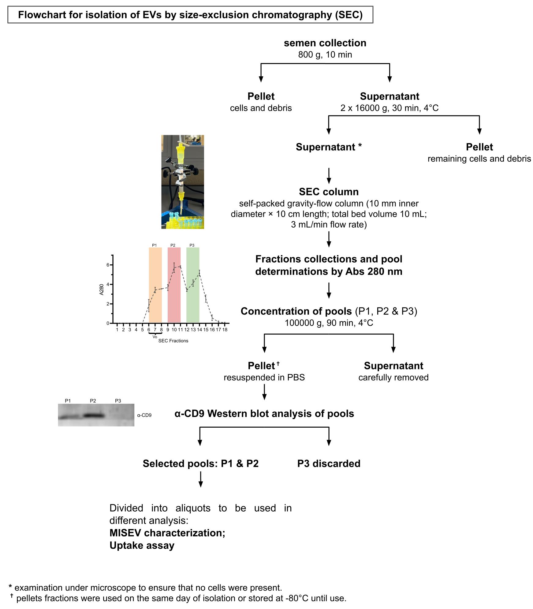
Figure 1. Flowchart for the isolation of extracellular vesicles (EVs) by size-exclusion chromatography (SEC). Seminal plasma is collected and subjected to differential centrifugation to remove cells and debris. The resulting supernatant is applied to a self-packed SEC column, and 18 consecutive fractions of 0.5 mL are collected. Protein content in each fraction is quantified by measuring absorbance at 280 nm (A280). Fractions corresponding to the peaks (P1–P3) are pooled and concentrated by ultracentrifugation at 100,000× g for 90 min at 4 °C. Pellets are resuspended in PBS and analyzed by western blot to assess the presence of the EV marker CD9. V0: void volume. P1–P3: pooled fractions. Representative images from independent replicates (n = 3) are shown. For a detailed overview of the column, see Supplementary Figure 1.
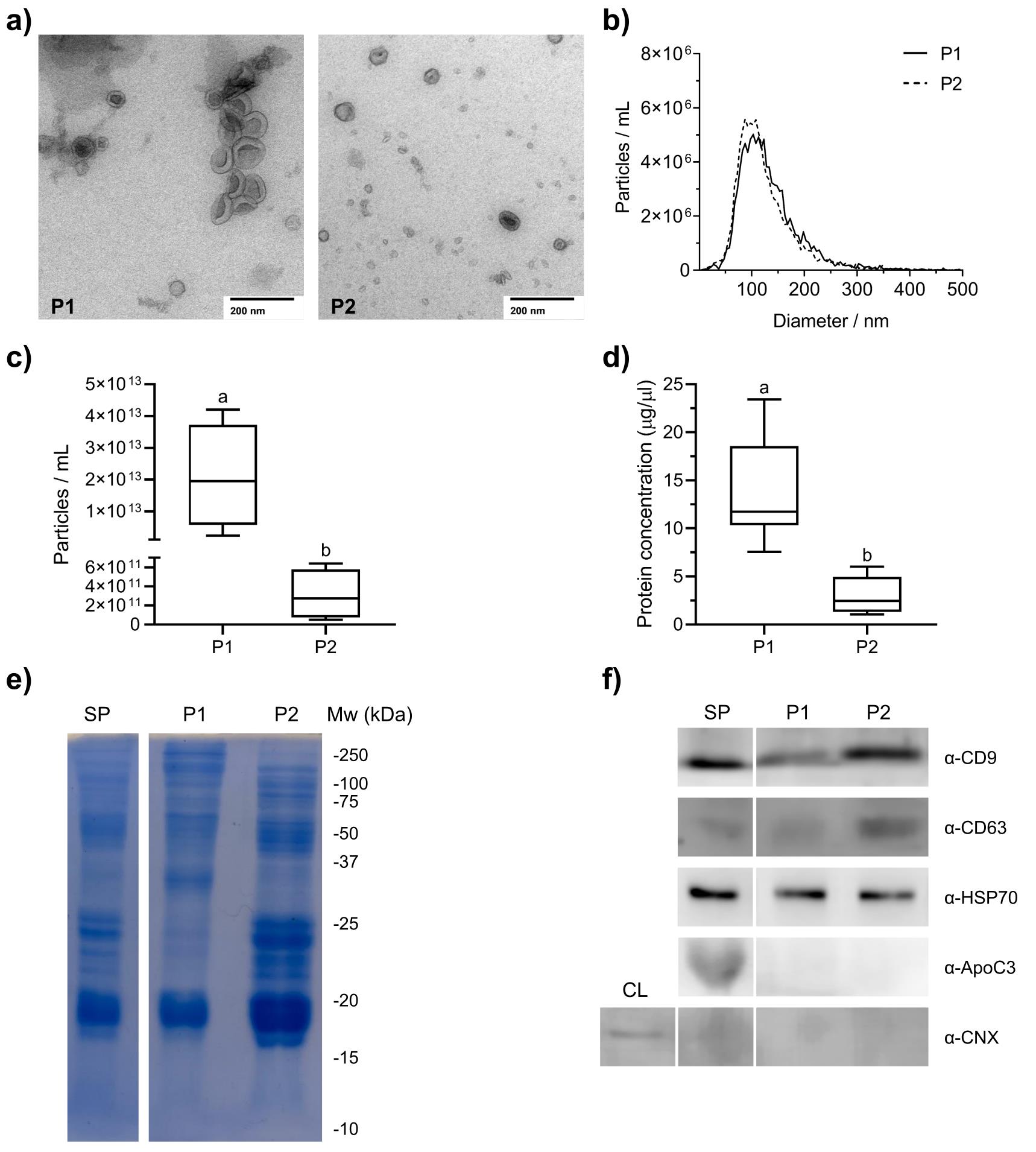
Figure 2. Characterization of seminal plasma (SP) extracellular vesicles (EVs). Analysis of SP-EVs fractions by (a) transmission electron microscopy (TEM) and (b, c) nanoparticle tracking analysis (NTA, n = 4). (d) Protein concentration (μg/μL) (n = 9) was assessed by Abs280 nm. (e) SDS-PAGE (15%) protein profile of SP-EV preparations and SP (60 μg protein/lane) (n = 3) stained with Coomassie brilliant blue. (f) Western blot analysis (60 μg protein/lane) of EV preparations and complete SP using antibodies against EV-associated proteins CD9, CD63, and HSP70, endoplasmic reticulum marker calnexin (CNX), and a non-vesicular extracellular particle apolipoprotein C-III (ApoC3) (n = 3) (full western blot images in Supplementary Figure 2). SP: seminal plasma; P1–2: pool of fractions in size-exclusion chromatography; CL: cell lysate; Mw: molecular weight marker. Different letters (a, b) above the bars indicate statistically significant differences (p < 0.05) between methods (C, P1, and P2), as determined by a linear mixed-effects model and Tukey-adjusted pairwise comparisons. A significant positive correlation was observed between particle concentration and protein concentration across samples (Spearman’s ρ = 0.747, p = 0.0009). All data are expressed as mean ± SD of four independent experiments (n = 4). Scale bar = 200 nm.
C. CFSE labeling of SP-EVs
1. Prepare a 5 mM CFSE (CellTrace CFSE Cell Proliferation kit) stock solution by adding 18 μL of the manufacturer-provided DMSO to the dye vial, following the reagent stock solution instructions supplied by the manufacturer.
2. Dilute the CFSE stock to a final concentration of 10 μM (manufacturer-recommended working concentration) in PBS containing EVs at a final protein concentration of 0.25 μg/μL. Include a dye-only control by substituting EVs with PBS to account for any residual CFSE that may co-precipitate during subsequent washing steps.
Critical: Ensure thorough mixing of the solution to achieve uniform labeling, as incomplete homogenization can lead to variability in fluorescence intensity across samples.
3. Incubate the mixture up to 60 min at room temperature (RT), protected from light.
4. Pellet the labeled EVs by ultracentrifugation at 100,000× g for 70 min at 4 °C. Resuspend the pellet in a volume of PBS not exceeding 200 μL.
5. Optional quality check: the incorporation of CFSE can be assessed using a fluorometer or any fluorescence-measuring instrument compatible with CFSE excitation/emission settings (492/517 nm).
D. Sperm preparation and incubation with labeled SP-EVs
1. Thaw semen straws in a water bath at 37 °C for 30 s. Dry straws to avoid osmotic shock and transfer the spermatozoa to prewarmed Eppendorf tubes at 37 °C.
2. Observe sperm subjective motility under a microscope.
3. For motile sperm selection, use the single-layer centrifugation (SLC) method using a single density solution, composed of colloidal silica coated with glycidoxypropyltrimethoxysilane (e.g., Ovipure® or Percoll) according to the manufacturer’s instructions [13]. Briefly, add two volumes of prewarmed Ovipure® density gradient to the spermatozoa and centrifuge at 800× g for 10 min. Recover the pellet (cloud at the bottom), which may vary in size depending on the initial sample.
4. To wash, add two volumes of prewarmed PBS to the pellet and centrifuge at 800× g for 5 min. Carefully remove the supernatant and recover the pellet. Work quickly to prevent spermatozoa from swimming back into the supernatant.
5. To measure concentration, dilute 5 μL of the recovered sample in 95 μL of water (1:20 dilution). Use a Neubauer chamber to count spermatozoa and determine concentration (cells/mL).
6. Incubate spermatozoa suspension with EVs at a ratio of 10 μg of EV protein per 1 million cells at 37 °C for up to 60 min. Make sure to include the following controls: spermatozoa without EVs (negative control); spermatozoa + free CFSE (control baseline of residual CFSE); and spermatozoa + CFSE 2 μM (as a positive control).
7. Analyze the CFESE-labeled sperm by flow cytometry and/or fluorescence microscopy (Figure 3). Ensure that all controls are properly set. These controls define the baseline CFSE fluorescence (grey histogram), which is used to set the CFSE- gate, whereas the positive control (green histogram) is included as a positive control and provides the upper limit for the CFSE+ gate. Together, these two controls establish the boundaries separating CFSE- and CFSE+ events. The experimental sperm samples incubated with CFSE-labeled EVs appear as black histograms. Calculate and compare the median fluorescence intensity of this distribution with the baseline. Any rightward shift of the black histogram relative to the dye-only baseline indicates uptake of CFSE-labeled EVs by spermatozoa. An example of these shifts and the corresponding quantification is shown in Figure 3.
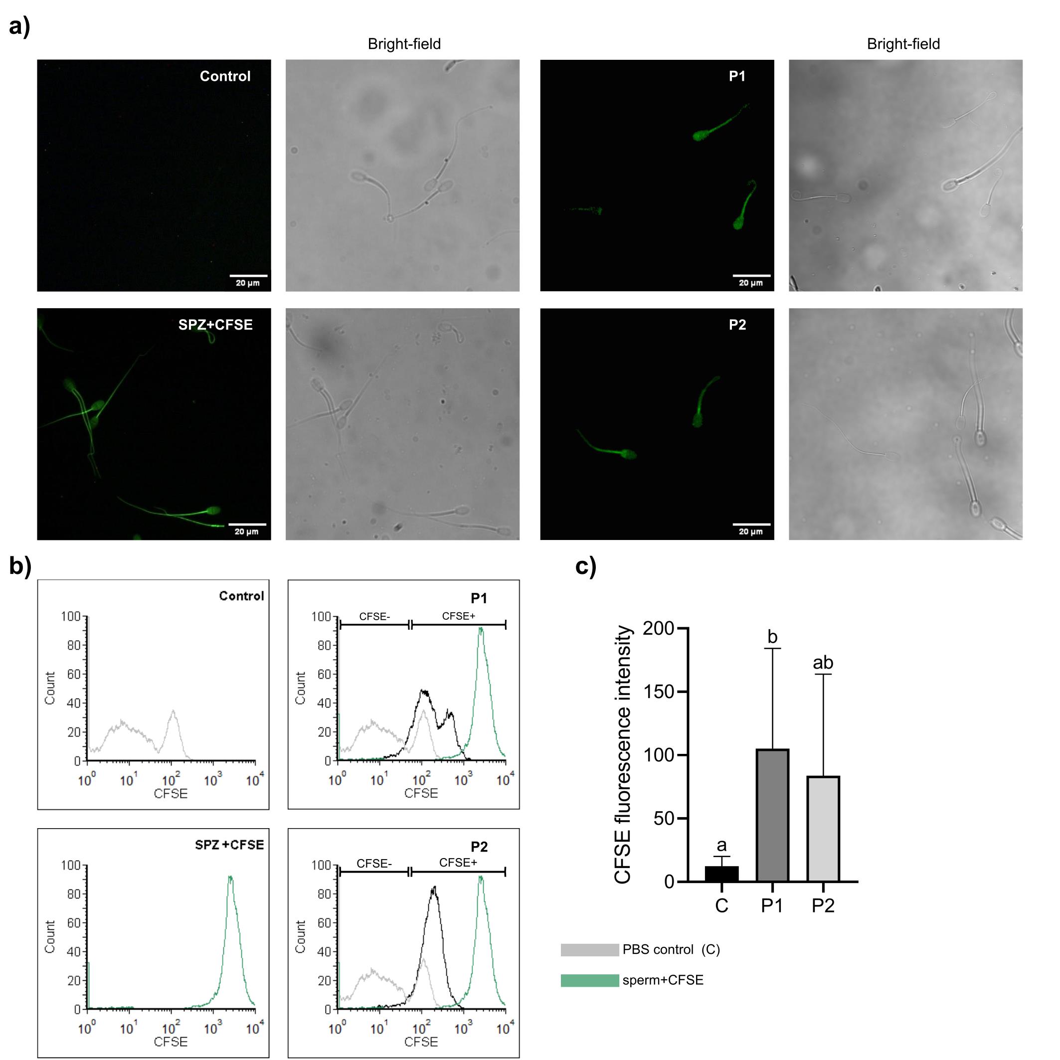
Figure 3. Uptake of labeled seminal plasma (SP) extracellular vesicles (EVs) by spermatozoa. Unlabeled spermatozoa were incubated with carboxyfluorescein succinimidyl ester (CFSE)-labeled SP-EVs, and fluorescence was assessed by flow cytometry (FL1 channel) and confocal microscopy. (a) Representative confocal images. (b) Flow cytometry histograms showing negative control (PBS + CFSE -no EVs-, gray line) and positive control (sperm + CFSE, green line) overlaid with experimental samples (black line). (c) Relative median ± SD fluorescence intensity of sperm cells incubated with CFSE-labeled EVs (n = 3). C: Negative control PBS + CFSE. P1 and P2: pool of fractions in SEC column. Different letters (a, b) indicate significant differences (p < 0.05) among treatments (C, P1, and P2) as determined by a linear mixed-effects model and Tukey-adjusted pairwise comparisons. Scale bar = 20 μm.
Data analysis
Data were analyzed using GLMM (generalized linear mixed-effects model) to determine the statistical significance between SP-EV isolation methods (explanatory variable) for each experiment. According to the experimental design, each seminal plasma sample (replicate) was considered as a random variable. All analyses were performed using R software.
A comparison of flow cytometry fluorescence intensity between treatments and the PBS control is available in [14] (see Methods section and Figure 3 of the referenced article). Briefly, after the incubation period, cells were washed and analyzed by flow cytometry (Partec, CyFlow Space, Germany). Data acquisition was performed using FloMax software, with a 488-nm argon-ion laser used to excite the probe. Emitted fluorescence was detected using the FL1 channel for CFSE (530/280 nm, excitation/emission). A dedicated acquisition template was set to distinguish spermatozoa from debris based on FSC vs. SSC. For each sample, a total of 10,000 spermatozoa were recorded, and subsequent data analysis was carried out using Flowing 2 software (University of Turku, Finland).
Validation of protocol
This protocol has been used and validated in the following research article:
Armani et al. [14]. Extracellular vesicles in ram seminal plasma: transport and transfer of capacitation regulating factors to sperm. Biol Reprod.
General notes and troubleshooting
General notes
1. This protocol can be applied to other experimental systems, taking into account possible sources of variation in the results.
2. The protocol can be scaled up by increasing the sample volume and proportionally adjusting the column dimensions. See Table 1.
3. Always follow the column manufacturer’s recommendations for packing, flow rates, and maximum pressure.
4. To preserve EV integrity, minimize shear, avoid drying the resin, and ensure cold conditions whenever feasible.
Table 1. Column scaling guidance
| Sample volume (mL) | Diameter (cm) | Height (cm) | Volume (mL) | Sample/column fraction |
|---|---|---|---|---|
| 0.4 | 0.7 | 7 | 10.78 | 3.71 |
| 0.4 | 1 | 10 | 31.42 | 1.27 |
| 0.8 | 0.7 | 14 | 21.55 | 3.71 |
| 0.8 | 1 | 20 | 62.83 | 1.27 |
| 1.2 | 0.7 | 21 | 32.33 | 3.71 |
| 1.2 | 1 | 30 | 94.25 | 1.27 |
| 1.2 | 1.5 | 45 | 318.09 | 0.38 |
| 2 | 0.7 | 35 | 53.88 | 3.71 |
| 2 | 1 | 50 | 157.08 | 1.27 |
Troubleshooting
Problem 1: Diluted EVs after ultracentrifugation.
Possible cause: Excess of buffer volume.
Solution: Resuspend the pellet with the minimum buffer volume and add more only if necessary.
Problem 2: Poor spermatozoa motility after thawing.
Possible causes: Improper thawing or osmotic shock.
Solution: Ensure semen straws are thawed quickly and dried properly before use.
Supplementary information
The following supporting information can be downloaded here:
1. Supplementary Figure 1. SuperdexTM 200 size-exclusion chromatography (SEC) column performance and fraction characterization.
2. Supplementary Figure 2. Full western blot images were obtained for the EV-specific markers CD9 (a), CD63 (b), HSP70 (c), and for the EV negative marker Calnexin (CNX) (d), and ApoC3 (e).
Acknowledgments
The authors would like to thank Viviana Daniel for her assistance with confocal microscopy and flow cytometry, and Dr. Gonzalo Caló for his support with ultracentrifugation procedures. We also thank Dr. Ana Josefa Soler Valls for providing reagents for the anti-CNX western blot, and Dr. Gabriela Berg for supplying reagents used in the anti-ApoC3 assay. This work was supported by grants from the National Scientific and Technical Research Council (CONICET, Argentina, PIP21-11220200100850CO awarded to A.C.) and the Society for the Study of Reproduction (2024 SSR Emerging Investigator Grant awarded to L.Z.). The methodology here is based on [15,16] and remains consistent with our previously reported protocol [14]. Author contributions were as follows: conceptualization, T.A., S.P.M., F.H., and A.C.; methodology, T.A., A.N., S.R., L.Z., J.L., and M.B.A.; investigation, formal analysis, validation, and visualization, all authors; writing—original draft preparation, T.A., A.N., S.R., L.Z., J.L., and M.B.A.; writing—review and editing, all authors; supervision, T.A., S.P.M., F.H., and A.C.; funding acquisition, A.C. and L.Z. All authors have read and approved the final version of the manuscript.
Competing interests
The authors declare that they have no competing interests.
Ethical considerations
All animals used in this study were managed in strict accordance with good animal practice and the conditions approved by the Animal Ethics Committee at INTA (Instituto Nacional de Tecnología Agropecuaria), Argentina (ID 217/2021; CICUAE INTA CeRBAS).
References
- van Niel, G., D'Angelo, G. and Raposo, G. (2018). Shedding light on the cell biology of extracellular vesicles. Nat Rev Mol Cell Biol. 19(4): 213–228. https://doi.org/10.1038/nrm.2017.125
- Margolis, L. and Sadovsky, Y. (2019). The biology of extracellular vesicles: The known unknowns. PLoS Biol. 17(7): e3000363. https://doi.org/10.1371/journal.pbio.3000363
- Jeppesen, D. K., Fenix, A. M., Franklin, J. L., Higginbotham, J. N., Zhang, Q., Zimmerman, L. J., Liebler, D. C., Ping, J., Liu, Q., Evans, R., et al. (2019). Reassessment of Exosome Composition. Cell. 177(2): 428–445.e18. https://doi.org/10.1016/j.cell.2019.02.029
- Franchi, A., Moreno‐Irusta, A., Domínguez, E. M., Adre, A. J. and Giojalas, L. C. (2019). Extracellular vesicles from oviductal isthmus and ampulla stimulate the induced acrosome reaction and signaling events associated with capacitation in bovine spermatozoa. J Cell Biochem. 121(4): 2877–2888. https://doi.org/10.1002/jcb.29522
- Xu, Z., Xie, Y., Zhou, C., Hu, Q., Gu, T., Yang, J., Zheng, E., Huang, S., Xu, Z., Cai, G., et al. (2020). Expression Pattern of Seminal Plasma Extracellular Vesicle Small RNAs in Boar Semen. Front Vet Sci. 7: e585276. https://doi.org/10.3389/fvets.2020.585276
- Barranco, I., Spinaci, M., Nesci, S., Mateo-Otero, Y., Baldassarro, V. A., Algieri, C., Bucci, D. and Roca, J. (2024). Seminal extracellular vesicles alter porcine in vitro fertilization outcome by modulating sperm metabolism. Theriogenology. 219: 167–179. https://doi.org/10.1016/j.theriogenology.2024.02.024
- Du, J., Shen, J., Wang, Y., Pan, C., Pang, W., Diao, H. and Dong, W. (2016). Boar seminal plasma exosomes maintain sperm function by infiltrating into the sperm membrane. Oncotarget. 7(37): 58832–58847. https://doi.org/10.18632/oncotarget.11315
- Lange-Consiglio, A., Capra, E., Monferini, N., Canesi, S., Bosi, G., Cretich, M., Frigerio, R., Galbiati, V., Bertuzzo, F., Cobalchini, F., et al. (2022). Extracellular vesicles from seminal plasma to improve fertilizing capacity of bulls. Reproduction and Fertility 3(4): 313–327. https://doi.org/10.1530/raf-22-0037
- Mehdinejadiani, S., Azad, N., Dehghan, Z., Khosravizadeh, Z., Saberi, F., Rezaee, D., Pilehchi, T., Goudarzi, N., Salahi, E., Mehdinejadiani, K., et al. (2025). Role of Seminal Exosomes in Reproduction. J Cell Physiol. 240(11): e70106. https://doi.org/10.1002/jcp.70106
- Welsh, J. A., Goberdhan, D. C. I., O'Driscoll, L., Buzas, E. I., Blenkiron, C., Bussolati, B., Cai, H., Di Vizio, D., Driedonks, T. A. P., Erdbrügger, U., et al. (2024). Minimal information for studies of extracellular vesicles (MISEV2023): From basic to advanced approaches. J Extracell Vesicles. 13(2): e12404. https://doi.org/10.1002/jev2.12404
- Konoshenko, M. Y., Lekchnov, E. A., Vlassov, A. V. and Laktionov, P. P. (2018). Isolation of Extracellular Vesicles: General Methodologies and Latest Trends. Biomed Res Int. 2018: 1–27. https://doi.org/10.1155/2018/8545347
- Cvjetkovic, A., Lötvall, J. and Lässer, C. (2014). The influence of rotor type and centrifugation time on the yield and purity of extracellular vesicles. J Extracell Vesicles. 3(1): e23111. https://doi.org/10.3402/jev.v3.23111
- Morrell, J. and Wallgren, M. (2011). Colloid Centrifugation of Boar Semen. Reprod Domest Anim. 46: 18–22. https://doi.org/10.1111/j.1439-0531.2011.01866.x
- Armani, T., Nicolli, A. R., Zalazar, L., Lobo, J. I., Arellano, M. B., Hozbor, F. A., Rio, S., Martinez, S. P. and Cesari, A. (2025). Extracellular vesicles in ram seminal plasma: transport and transfer of capacitation regulating factors to sperm. Biol Reprod. 114(2): 555–569. https://doi.org/10.1093/biolre/ioaf248
- Yang N, Zhao C, Kong L, Zhang B, Han C, Zhang Y, Qian X, Qin W. (2023). Absolute Quantification of Dynamic Cellular Uptake of Small Extracellular Vesicles via Lanthanide Element Labeling and ICP–MS. Anal Chem. 95:11934–11942. https:/doi.org/10.1021/acs.analchem.3c01421
- Wang, H., Zhu, Y., Tang, C., Zhou, Z., Wang, Z., Li, Z., Zheng, X., Chen, S., Zhou, Y., Liang, A., et al. (2021). Reassessment of the Proteomic Composition and Function of Extracellular Vesicles in the Seminal Plasma. Endocrinology. 163(1): e1210/endocr/bqab214. https://doi.org/10.1210/endocr/bqab214
Article Information
Publication history
Received: Jan 10, 2026
Accepted: Mar 2, 2026
Available online: Mar 26, 2026
Published: Apr 5, 2026
Copyright
© 2026 The Author(s); This is an open access article under the CC BY-NC license (https://creativecommons.org/licenses/by-nc/4.0/).
How to cite
Armani, T., Nicolli, A. R., Zalazar, L., Lobo, J. I., Arellano, M. B., Hozbor, F. A., Rio, S., Martinez, S. P. and Cesari, A. (2026). Uptake Assay of Ram Seminal Plasma Extracellular Vesicles to Sperm. Bio-protocol 16(7): e5653. DOI: 10.21769/BioProtoc.5653.

 Download Citation in RIS Format
Category
Developmental Biology > Reproduction
Cell Biology > Organelle isolation > Extracellular vesicle
Cell Biology > Cell-based analysis > Cell-to-cell interaction
Do you have any questions about this protocol?
Post your question to gather feedback from the community. We will also invite the authors of this article to respond.
 Post a Question


