

Evaluating Thioredoxin-Mediated CFoCF1 Reduction Using an In Vitro Thylakoid Assay
利用体外类囊体实验评估硫氧还蛋白介导的 CFoCF1 还原
The activity of chloroplast ATP synthase (CFoCF1) is precisely regulated through a thioredoxin (Trx)-mediated dithiol/disulfide reaction in response to varying light conditions. This regulatory mechanism is further controlled by ΔpH formation across the thylakoid membrane. To better understand this complicating regulatory function of CFoCF1, a method is required to evaluate the extent of CFoCF1 reduction by Trx under controlled ΔpH conditions and to directly evaluate the redox state of CFoCF1. In this study, we present a simple in vitro procedure to assess the CFoCF1 reduction system using spinach thylakoids. The method consists of three key steps: (A) simple preparation of intact thylakoids from spinach leaves; (B) reduction of CFoCF1 on the thylakoid membrane using recombinant Trx under light irradiation; and (C) in situ determination of the redox state of CFoCF1 by labeling thiol groups with a maleimide reagent followed by protein detection using western blotting. The redox state of CFoCF1 was determined by mobility shifts on non-reducing SDS-PAGE. This protocol provides a refined strategy for elucidating the regulatory mechanism controlling energy conversion by CFoCF1 under fluctuating photosynthetic conditions.
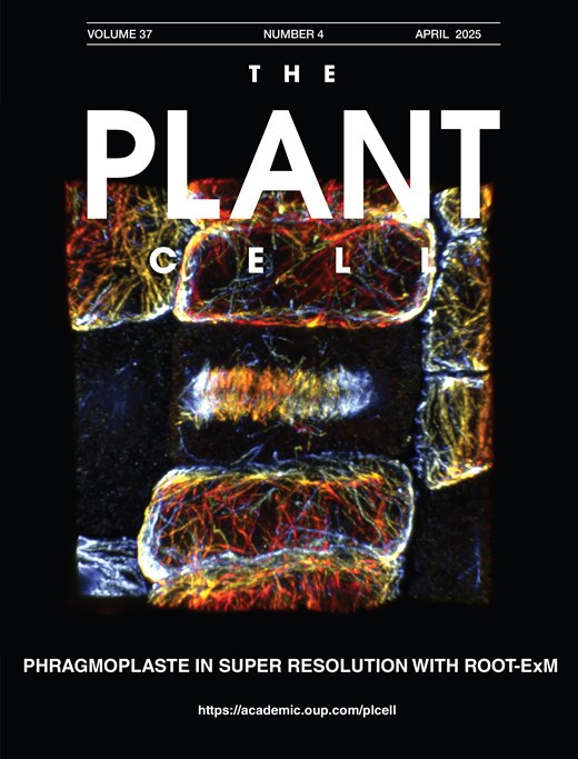
ROOT-ExM: Super-Resolution Imaging of Proteins in Arabidopsis Roots by Expansion Microscopy
ROOT-ExM:利用膨胀显微技术实现拟南芥根部蛋白质的超分辨率成像

Conventional light microscopy is limited in resolution by the diffraction limit of light, restricting the visualization of the nanoscale organization of biomolecules. Expansion microscopy (ExM) has emerged as a powerful technique to overcome this barrier by physically expanding the specimen embedded in a swellable hydrogel without requiring specialized or high-cost imaging hardware. ExM is widely used in animal models, whereas its application to plant tissues has been challenging due to their multicellularity, in which each cell is encompassed by the rigid cell wall, which resists the expansion forces and prevents isotropic swelling. Here, we describe a robust and optimized ExM protocol specifically designed for Arabidopsis thaliana root tissues. This protocol details critical steps, including immunostaining, anchoring, gelation, denaturation, cell wall digestion, and expansion. Our method achieves an expansion factor of approximately 4.3×, enabling an effective lateral resolution of ~60 nm using a standard confocal microscope. We demonstrate the visualization of microtubules with preserved ultrastructure. This accessible protocol allows plant researchers to perform super-resolution imaging without specialized optical equipment, facilitating detailed structural analysis of plant cells.
Biolayer Interferometry (BLI) to Quantify RALF1–Pectin Interactions
利用生物层干涉技术定量分析 RALF1 与果胶的相互作用
Cellular function relies on a network of precisely regulated interactions among macromolecules such as proteins, peptides, carbohydrates, and nucleic acids. These molecular interactions regulate vital processes, including signaling, structural organization, and developmental patterning. Biolayer interferometry (BLI) is a label-free optical biosensing technique that enables real-time quantification of such interactions. This protocol describes how to use BLI to assess the binding affinity between a biotinylated plant peptide hormone (RALF1) and cell wall–derived oligogalacturonides (OG25–50) on the Octet RED96 platform. Streptavidin-coated biosensors are employed to immobilize the ligand, while analyte binding is monitored through wavelength shifts in the reflected light. The protocol includes detailed steps for sensor preparation, assay setup, software configuration, and kinetic data analysis. While optimized for plant peptide–matrix interactions, the method is broadly adaptable to other macromolecular systems across biological disciplines.
Analysis of Cauline Leaf Development in Arabidopsis thaliana Using Time-Lapse Confocal Microscopy
利用延时共聚焦显微成像分析拟南芥茎生叶发育过程
Understanding cellular growth dynamics in plants requires precise, long-term imaging of developing tissues. Cauline leaves are produced during the transition from vegetative to reproductive development and provide a useful system for studying how laminar organs diversify in form and function. While other laminar organs, such as rosette leaves and sepals, have been extensively studied, early cauline leaf development remains technically challenging to capture due to their concealed position, curved morphology, and the presence of dense trichomes. Here, we provide a complete pipeline for the dissection, confocal imaging, 2.5D segmentation, and image analysis of initiating cauline leaves in Arabidopsis thaliana. This method enables reproducible, high-resolution imaging of cauline leaves, supporting robust quantitative analysis of growth across developmental stages at cellular scale resolution.
3D Reconstruction of Mature Arabidopsis Ovules Using FIB-SEM to Study Filiform Apparatus Morphology
利用 FIB-SEM 三维重建成熟拟南芥胚珠以研究丝状器形态
Volume electron microscopy based on serial sectioning allows for three-dimensional (3D) visualization and analysis of the internal structures of tissues, cells, and organelles. One such technique, focused ion beam (FIB) scanning electron microscopy (SEM), has the advantages of nanoscale sectioning and high z-resolution, but the disadvantage of limited volume processing. Because of this limitation, targeting localized objects by FIB-SEM is difficult. Here, we developed a FIB-SEM observation workflow that enables the analysis of the filiform apparatus of synergid cells enclosed in the Arabidopsis ovule. In this protocol, plant samples are stained, embedded, trimmed, and carbon-coated while maintaining their orientation within the tissue. Then, sequential observations are performed using Cut & See function of FIB-SEM, followed by image processing for 3D reconstruction. Utilization of multi-scanning and image cropping from high-resolution data helps to identify localized targets within plant tissue. The filiform apparatus, which is an invaginated cell wall structure of the synergid cells, shows distinct contrast in each image, allowing for segmentation using brightness-based binarization. Such segmentation avoids the need to manually trace complex structures and facilitates 3D reconstruction by volume electron microscopy.
Quantitative Assessment of Heat Shock-Induced Ferroptosis-Like Cell Death via Electrolyte Leakage in Arabidopsis thaliana Seedlings
拟南芥幼苗热激诱导铁死亡样细胞死亡的电解质渗漏定量评估
We present a protocol to allow continuous assessment of cell death in Arabidopsis thaliana (L.) seedlings by measuring the release of electrolytes from dying cells upon heat shock. The electrolyte leakage assay is a well-established method to quantify the extent of cell death of plant tissues exposed to pathogen infection, since the activation of the immune response leads to compromised membrane integrity and to the release of ions from the dying cell. This prolonged release of electrolytes is considered a hallmark of regulated cell death in plants. Heat shock in plants induces ferroptosis-like cell death, which can be suppressed either pharmacologically, using inhibitors such as ferrostatin, or genetically through knockout of ferroptosis-related genes. Here, we have adapted the electrolyte leakage assay to quantify cell death in young Arabidopsis seedlings exposed to a heat shock previously shown to induce ferroptosis-like cell death. We also illustrate how this method can be used to assess activation of ferroptosis-like cell death in whole Arabidopsis seedlings using ferrostatin or knockout mutants of potential gene candidates involved in ferroptosis-like cell death.
TALENs and Related Technologies for Editing Nuclear and Organellar Genomes in a Model Plant, Arabidopsis thaliana
TALENs及相关技术在模式植物拟南芥核基因组与细胞器基因组编辑中的应用
Plant genome editing is a powerful approach for modifying plant DNA to investigate gene function and to engineer desirable traits. Several genome-editing technologies have been developed, among which CRISPR/Cas systems and transcription activator-like effector nucleases (TALENs) are widely used to introduce targeted double-stranded DNA breaks. While CRISPR/Cas systems are highly efficient for nuclear genome editing, their application to plant organellar genomes remains limited, largely due to difficulties in guide RNA delivery into mitochondria and chloroplasts. Here, we present a detailed and reproducible protocol for constructing TALEN-based binary vectors for targeted genome editing in Arabidopsis thaliana. This protocol describes the assembly of TALE repeat arrays, the generation of nuclear-, mitochondrial-, and plastid-targeted TALEN expression vectors using MultiSite Gateway cloning, and subsequent Agrobacterium-mediated plant transformation and genotyping. The workflow enables the production of nTALENs, mitoTALENs, and ptpTALENs using a unified vector design strategy. In addition, the protocol briefly outlines the construction principles of TALE-based cytidine deaminases (TALECDs) for targeted C-to-T base editing in plant organellar genomes. The protocol provides a flexible and robust framework for plant nuclear and organellar genome editing and can be readily adapted to different target genes and experimental purposes. Its modular design and compatibility with standard molecular cloning techniques make it accessible to laboratories aiming to perform precise genome manipulation in plants.
Kinetic Determination of Cytochrome b6f Activity In Vitro
体外动力学测定细胞色素 b6f 复合体活性
While traditional kinetic studies of the cytochrome b6f complex have frequently relied on measurements within the complex environment of intact leaves or whole-organism systems, such approaches can be limited by overlapping signals and physiological variables. This protocol advances existing frameworks by introducing a streamlined, multi-wavelength spectroscopic approach utilizing a reconstituted in vitro system to elucidate the inter-complex electron transfer kinetics between photosystem I and cytochrome b6f. Utilizing the JTS-150 pulsed spectrometer, supplied with a Smart Lamp, we monitored the redox transitions of P700+ and Cytf by simultaneously measuring the absorbance changes of our isolated complexes system in six different wavelengths (546, 554, 563, 574, 705, and 740 nm). Kinetic analysis was divided into two phases: laser-induced flash kinetics and steady-state actinic induction. We resolved the second-order re-reduction of P700+ by plastocyanin, accounting for detector saturation constraints with a 2 ms post-flash delay. Steady-state measurements under actinic light revealed complex Cytf turnover, characterized by a double-exponential decay. Furthermore, dark relaxation kinetics were used to quantify ferredoxin-mediated re-reduction of the cytochrome pool. By allowing the incorporation of specific regulatory and inhibitory factors, this methodology sets the ground for the deconvolution of competing electron pathways. It can therefore be used as a robust framework for assessing the mechanism of regulatory processes on photosynthetic flux.
Detecting Touch-Induced Calcium Dynamics With Live-Cell Imaging in Torenia Stigma
利用活细胞成像检测蓝猪耳柱头中的触碰诱导钙动态变化
Calcium ions serve as a universal secondary messenger, integrating diverse external signals, such as light, herbivory, and mechanical stimuli, within plant cells. However, the visualization and mechanistic dissection of calcium signaling specifically in response to mechanical stimulation remain technically challenging and underexplored in most plants. Previous studies have been largely confined to a few model systems, including Arabidopsis; here, we introduce a live-cell imaging approach using the stigmas of Torenia fournieri. This in vitro system enables multiscale observation of calcium signal patterns following controlled mechanical stimulation. This versatile platform not only simplifies the design of calcium imaging assays but also provides a tractable system for functionally validating other key molecular components in this signaling pathway.
Extraction and Isolation of Extracellular Vesicles From Piper betle Leaves Using the Apoplastic Fluid Washing and Size Exclusion Chromatography Method
基于质外体液洗脱与尺寸排阻色谱法提取和分离蒌叶来源的细胞外囊泡

Plant-derived extracellular vesicles (PDEVs) have emerged as important mediators of intercellular communication and hold growing potential in therapeutic applications. However, standardized methods for their isolation, particularly from Piper betle leaves (PBL), remain unexplored. Existing apoplastic fluid washing (AFW) extraction techniques typically rely on manual syringe infiltration, which often leads to inconsistent pressure control, variable yields, and increased risk of tissue damage. This protocol describes a vacuum-assisted AFW extraction method optimized for the recovery of intact extracellular vesicles (EVs) from PBL. The workflow features controlled negative pressure using a vacuum pump and chamber to achieve more efficient leaf infiltration compared to infiltration using the syringe method and reproducible apoplastic fluid (AF) collection with subsequent low-speed centrifugation steps, to ensure minimal contamination and preservation of vesicle integrity. Piper betle–derived extracellular vesicle (PBdEV) isolation and purification steps are performed using size exclusion chromatography (SEC). The size and concentration of PBdEVs were confirmed using nanoparticle tracking analysis (NTA), whereas the cup-shaped and lipid bilayer morphology of the EVs were confirmed using transmission electron microscopy (TEM). The method is scalable and adaptable to various leaf morphologies and physiological states, making it suitable for both exploratory and high-throughput studies. Overall, this protocol provides a more consistent, efficient, and tissue-preserving alternative to traditional syringe-based AF extraction methods, offering higher-quality EV preparations for plant EV research.
Spatial Imaging and Quantification of Hydrogen Peroxide in Arabidopsis Roots: From Sample Preparation to Image Analysis
拟南芥根中过氧化氢的空间定位成像及定量分析:从样品制备到图像分析
Reactive oxygen species (ROS) are central regulators of plant development and stress responses, with hydrogen peroxide (H2O2) acting as a key signaling molecule whose spatial distribution determines adaptive versus damaging outcomes. Accurate detection of H2O2 at tissue and cellular resolution is therefore essential for understanding redox-dependent regulation of plant growth. A variety of techniques have been used to monitor H2O2, including bulk spectrophotometric and fluorometric assays, genetically encoded sensors for real-time measurements, and chemical probes for in situ detection. While these approaches differ in sensitivity, specificity, and temporal resolution, many are limited by a lack of spatial information, technical complexity, or dependence on transgenic material. Here, we present a detailed protocol for 3,3′-diaminobenzidine (DAB)-based histochemical detection of H2O2 in seedling roots, covering staining, imaging, and semi-quantitative image analysis using open-source software (FIJI/ImageJ). The method relies on peroxidase-mediated oxidation of DAB, resulting in a stable, light-resistant, and insoluble precipitate that enables visualization of H2O2 accumulation with high spatial resolution. This protocol provides a robust, accessible, and genetically independent approach for spatial analysis of H2O2 in plant tissues. Its simplicity, compatibility with diverse genotypes and treatments, and suitability for semi-quantitative analysis make it a valuable tool for examining the spatial distribution of H2O2, thereby providing spatial insight into redox-related regulatory processes during plant development and stress responses.
A Step-by-Step Protocol for the Isolation of Aloe vera–Derived Extracellular Vesicles via Manual and Shear-Force Homogenization
采用手工匀浆与剪切力匀浆法分离芦荟来源细胞外囊泡的逐步实验方案
Aloe vera has long been used for its diverse pharmacological properties, motivating continued interest in isolating and preserving the bioactive molecules responsible for its therapeutic potential. More recently, Aloe vera–derived extracellular vesicles (Av-EVs) have emerged as nanoscale, cell-free carriers capable of retaining and delivering these properties, making them attractive for various biomaterials, nanomedicine, and regenerative medicine applications. Multiple techniques are available for extracellular vesicle isolation. These include ultracentrifugation, polymer-based precipitation, size-exclusion chromatography, immunoaffinity capture, ultrafiltration, density gradient separation, and emerging microfluidic platforms. Each method presents distinct trade-offs in purity, yield, scalability, and downstream compatibility. Despite this diversity, standardized workflows tailored to Av-EV isolation remain limited, and the influence of homogenization-induced shear forces and plant maturity on vesicle recovery and characterization has not been systematically addressed. Here, we present a reproducible protocol for isolating Av-EVs from Aloe vera gel employing two distinct homogenization strategies: manual, no-shear force (NB EVs), and blender-based shear-force homogenization (B EVs). The workflow covers gel preparation, serial centrifugation for debris removal, ultracentrifugation as the gold standard for vesicle enrichment, and final sterile filtration. This protocol enables consistent recovery of Av-EVs suitable for physicochemical characterization and functional analyses. It is simple and relies on commonly available laboratory equipment, facilitating broad adoption by ultracentrifugation users and offering adaptability to diverse research projects involving purified Aloe vera gel and Av-EVs, including studies focused on wound healing, fibrotic scarring, and regenerative processes, where coordinated antioxidant, anti-inflammatory, antimicrobial, immunomodulatory, and moisturizing responses are of interest.
- 1
- 2
- 3
- 4
- 5
- 6
- 90