

In-Culture Antibody Capture Using Transient CHO Expression Systems
利用瞬时 CHO 表达系统在培养中捕获抗体
Antibody therapeutics have demonstrated transformative impacts on improving the quality of life of millions of patients, whereas advances in antibody discovery technologies have imposed a significant production challenge for the generation of a large diversity of therapeutic antibody candidates. A demand for the rapid production of dozens of purified antibodies in 10-mg quantities is entailed for functional screening and molecular assessment studies. Here, we present a robust semi-automated production protocol that bridges the gap between miniaturized high-throughput screenings and conventional custom-scale workflows. This methodology and workflow utilize a simple high-titer transient Chinese hamster ovary (CHO) cell host–CHO4Tx® expression system, a procedure of magnetic protein-A bead in-culture antibody capturing, and a semi-automated purification process with the GenScript AmMagTM SA Plus system. This production protocol has been proven to be robust and valuable for the routine production of dozens of antibody constructs per week in sufficient quality and quantity for cell-based and biophysical studies.
Comprehensive Protocol for Handling Human Small Airway Epithelial Cells (HSAECs) to Establish Air–Liquid Interface (ALI) Cultures With TEER-Based Barrier Integrity Assessment
人小气道上皮细胞气液界面培养及基于 TEER 的屏障完整性评估操作方案
Understanding epithelial barrier function is essential for studying both its normal physiology and its role in disease, yet choosing an appropriate experimental model remains challenging. Animal models are commonly used but often suffer from interspecies differences that limit translational relevance. Human-derived cell lines offer a more suitable alternative, although establishing them often requires immortalisation strategies that involve overexpression of oncogenes, which can introduce phenotypic and functional changes. In contrast, primary cells, such as human small airway epithelial cells (HSAECs), provide a more physiologically accurate model. A critical aspect of replicating the native respiratory environment is maintaining continuous air exposure, which can be achieved through air–liquid interface (ALI) culture. This protocol provides a unified, step-by-step workflow for cultivating primary HSAECs under ALI conditions, covering the entire process from initial recovery after cryopreservation to the formation of a barrier-like layer. The protocol incorporates non-invasive methods such as transepithelial electrical resistance (TEER) measurements to monitor its integrity. While individual elements of this workflow have been described separately in different studies, a consolidated version encompassing the full workflow has not been widely available. This resource is intended for researchers with limited experience in airway epithelial culture and offers practical, clear guidance through each step of the process.

Oligo(dT) Fluorescence In Situ Hybridization to Visualize the Poly(A) mRNAs in the Internal Tissues of Drosophila
利用 Oligo(dT) 荧光原位杂交观察果蝇内部组织中的 poly(A) mRNA

Fluorescence in situ hybridization (FISH) is a cytological method used to visualize specific oligonucleotide sequences within the cell. This method relies on the specific binding of a fluorescence-tagged probe, a short stretch of single-stranded polynucleotide, to its complementary sequence in the DNA or RNA, forming stable double-stranded hybrids. Fluorochromes, such as fluorescein, Alexa Fluor, cyanine dyes, or rhodamine, are attached to these probes to help in detecting their presence within the cell. Based on sequence complementarity, FISH allows for the visualization of the DNA or RNA with which they have hybridized. The distribution of these fluorochrome-tagged probes can be observed under a fluorescence or confocal microscope. The oligo(dT) FISH technique specifically utilizes a fluorochrome-tagged stretch of 40–50 thymidine (T) oligonucleotides that binds to the poly(A) tails of mature mRNAs within the cell. Newly transcribing pre-mRNAs and certain non-coding RNAs may not have poly(A) tails and therefore cannot be detected by this method. This step-by-step protocol outlines the oligo(dT) FISH technique for visualizing the cellular distribution of polyadenylated mRNAs in the tissues of Drosophila and other related model organisms.

Histomorphometrical Analyses of the Mouse Suprachiasmatic Nucleus
小鼠视交叉上核的组织形态计量学分析
The mammalian central circadian clock resides in the suprachiasmatic nucleus (SCN) of the hypothalamus in the brain and is responsible for coordinating daily rhythms of biological processes spanning from gene expression to behavior. Light, the primary environmental zeitgeber, entrains the SCN via melanopsin-expressing intrinsically photosensitive retinal ganglion cells that project through the retino-hypothalamic tract. Altered circadian rhythms are common in individuals diagnosed with neurodevelopmental and neurodegenerative disorders, and often, associated with structural alterations of the SCN and impaired retinal input; importantly, these anomalies can be recapitulated in animal models. Here, we describe step-by-step protocols for quantitative histomorphometrical analysis of the SCN and the assessment of retinal–SCN connectivity, previously used in mouse models of neurodevelopmental and neurodegenerative disorders. These include measurement of the SCN area, perimeter, height and width using Nissl- or DAPI-stained coronal sections, as well as densitometric and plot profile analyses of cholera toxin β-subunit–labeled retinal projections using Axiovision or Fiji/ImageJ. The protocols incorporate standardized region-of-interest, measurements by masked observers, and consistent scaling procedures to enhance reproducibility. These methods provide a rigorous framework for detecting structural anomalies and connectivity defects in the circadian system and can be broadly applied to other experimental models of circadian dysfunction.

Computational Quantification of Mouse Retinal Vasculature Using ImageJ
基于 ImageJ 的小鼠视网膜血管图像定量分析
Postnatal mouse retinal vascular development is a widely used model for studying retinal vascular diseases and evaluating candidate therapies. This is particularly relevant for inherited disorders such as familial exudative vitreoretinopathy (FEVR), in which impaired vascular growth and organization are central to disease pathogenesis. Numerous approaches have been used to assess retinal vasculature in mouse flat mounts, ranging from qualitative descriptions to limited quantitative measurements of vascular growth. However, phenotypic variability across genetic models, including different models of FEVR, complicates comparisons and underscores the need for standardized, comprehensive multi-parameter analyses that are suitable for rapid and cost-effective screening studies. We describe a standardized morphometric protocol using ImageJ software to quantitatively analyze mouse retinal vasculature in a reproducible manner. The protocol begins with measurement of areas of vascular disorganization (meshes) as well as total vascular and retinal area. Two defined regions in the peripheral and midperipheral retina are then selected to quantify cell clusters, followed by image processing, binarization, and skeletonization. From these processed images, vascular density, branch number, branch length and thickness, junction number, triple points, and box-counting fractal dimension and lacunarity are quantified. Overall, this protocol provides a rapid, cost-effective, and standardized framework for quantifying retinal vascular phenotypes across diverse mouse models. By capturing multiple structural features and accommodating phenotypic variability, it is well-suited for comparative studies and therapeutic screening in retinal vascular disease.
Evaluating Thioredoxin-Mediated CFoCF1 Reduction Using an In Vitro Thylakoid Assay
利用体外类囊体实验评估硫氧还蛋白介导的 CFoCF1 还原
The activity of chloroplast ATP synthase (CFoCF1) is precisely regulated through a thioredoxin (Trx)-mediated dithiol/disulfide reaction in response to varying light conditions. This regulatory mechanism is further controlled by ΔpH formation across the thylakoid membrane. To better understand this complicating regulatory function of CFoCF1, a method is required to evaluate the extent of CFoCF1 reduction by Trx under controlled ΔpH conditions and to directly evaluate the redox state of CFoCF1. In this study, we present a simple in vitro procedure to assess the CFoCF1 reduction system using spinach thylakoids. The method consists of three key steps: (A) simple preparation of intact thylakoids from spinach leaves; (B) reduction of CFoCF1 on the thylakoid membrane using recombinant Trx under light irradiation; and (C) in situ determination of the redox state of CFoCF1 by labeling thiol groups with a maleimide reagent followed by protein detection using western blotting. The redox state of CFoCF1 was determined by mobility shifts on non-reducing SDS-PAGE. This protocol provides a refined strategy for elucidating the regulatory mechanism controlling energy conversion by CFoCF1 under fluctuating photosynthetic conditions.
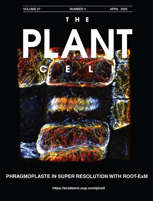
ROOT-ExM: Super-Resolution Imaging of Proteins in Arabidopsis Roots by Expansion Microscopy
ROOT-ExM:利用膨胀显微技术实现拟南芥根部蛋白质的超分辨率成像

Conventional light microscopy is limited in resolution by the diffraction limit of light, restricting the visualization of the nanoscale organization of biomolecules. Expansion microscopy (ExM) has emerged as a powerful technique to overcome this barrier by physically expanding the specimen embedded in a swellable hydrogel without requiring specialized or high-cost imaging hardware. ExM is widely used in animal models, whereas its application to plant tissues has been challenging due to their multicellularity, in which each cell is encompassed by the rigid cell wall, which resists the expansion forces and prevents isotropic swelling. Here, we describe a robust and optimized ExM protocol specifically designed for Arabidopsis thaliana root tissues. This protocol details critical steps, including immunostaining, anchoring, gelation, denaturation, cell wall digestion, and expansion. Our method achieves an expansion factor of approximately 4.3×, enabling an effective lateral resolution of ~60 nm using a standard confocal microscope. We demonstrate the visualization of microtubules with preserved ultrastructure. This accessible protocol allows plant researchers to perform super-resolution imaging without specialized optical equipment, facilitating detailed structural analysis of plant cells.

Measuring Electrophysiological Activity in Acute Brain Slices, Spheroids, and Organoids Using 3D High-Density Multielectrode Arrays
利用三维高密度多电极阵列检测急性脑片、脑球体和脑类器官的电生理活动
Animal and human stem cell–derived three-dimensional models to study physio-pathological brain functioning are becoming a gold standard for in vitro electrophysiology, as they enable the recapitulation of complex network properties by accounting for spatial architectural features that better reflect in vivo conditions than simpler 2D models. Standard planar multielectrode arrays (MEAs), typically providing tens of recording electrodes, are commonly used to record activity from 2D neuronal cultures. However, when adapted for use with 3D models, planar 2D MEAs showed limited effectiveness. The main issues are limited specimen adhesion to the chip, a low number of sensing elements, inability to retrieve signals from within the tissue, and reduced perfusion and vitality of the tissue in contact with sensors. To overcome these limitations, a new generation of microchip-based 3D high-density MEAs (3D HD-MEA) has been developed and validated in recent years. This technological advancement has improved the sensing capabilities and the vitality of 3D models, providing a tool tailored to maximize their potential. Here, we present an optimized protocol for neural network activity recordings in 3D models (including acute slices, brain spheroids, and organoids) from various brain regions using 3D HD-MEAs. First, we summarize the critical steps for 1) obtaining viable acute slices from the mouse cerebellum, cortico-hippocampal circuit, and prefrontal cortex, 2) establishing efficient coupling of the slices with the chip, and 3) performing recordings and analyses. We then describe the main procedures required to obtain human and animal brain spheroids and neural organoids, as well as standardized routines to perform effective recordings and analyses. For each section, we highlight the crucial steps, identify tips for specific applications, and propose troubleshooting procedures. For example, the same type of preparation (e.g., acute slices) requires different adjustments when working with different brain areas. The specific information provided here is intended to assist researchers in their daily efforts to obtain efficient and reproducible functional recordings from 3D models by using the cutting-edge technique of 3D HD-MEA.
Versatile Dual Mounting Enables Larval Zebrafish Imaging Across Microscope Configurations
双向固定方法支持斑马鱼幼体在不同显微镜配置下成像
Larval zebrafish are often mounted laterally to ensure consistent anatomical positioning and to standardize imaging of body axes across early development. However, this conventional approach often tethers sample orientation to a single microscope configuration and limits optical accessibility. We present a mounting protocol for larval zebrafish that enables optical access from both dorsal and ventral orientations while preserving lateral sample position. This approach uses common laboratory consumables to establish a mounting platform that eliminates any need to remount samples between the use of upright and inverted microscopes. By establishing a hydrophobic seal, mounted embryos can be inverted with ease to access the sample from either orientation. A seamless transition here facilitates reliable identification and longitudinal tracking of the same biological region of interest across microscope configurations. This protocol is broadly applicable to live imaging experiments requiring flexibility in imaging geometry, minimal sample handling, and high reproducibility.
Quantitative Analysis of Cell and Tissue Shape During Mouse Cranial Neural Tube Closure
小鼠颅部神经管闭合过程中细胞与组织形态的定量分析
Neural tube closure is a critical process that transforms the neural plate, an open epithelial tissue, into the closed tube that serves as the structural basis of the central nervous system. Defects in this process are among the most common and severe developmental diseases in the human population, with failures in cranial closure accounting for approximately one-third of total defects. However, the cell and tissue mechanisms that drive cranial closure remain opaque relative to the better studied process of spinal closure, in large part due to the unique challenges in characterizing cranial tissues. Here, we present protocols for quantifying cell dynamics and tissue-level remodeling events that enable highly spatiotemporally resolved investigations of the causes of cranial closure defects in mouse embryos. These include brightfield morphometric approaches, fluorescent staining and confocal imaging, and quantitative pipelines to analyze these image-based datasets. At the conclusion of these approaches, users will be able to quantify several parameters of overall tissue shape in the cranial neural tissues and produce rich quantitative datasets about cell-level parameters, particularly apical cell area. These can be used to identify correlative and causative differences between mutants and control embryos. Given their flexibility, many of these approaches can be generalized to other tissue morphogenetic contexts.

Detection of Target Molecules Within One-Millimeter-Thick Mouse Brain Slices by Using Peroxidase-Fused Nanobodies and Fluorochromized Tyramide-Glucose Oxidase Reaction
利用过氧化物酶融合纳米抗体和荧光化酪酰胺-葡萄糖氧化酶反应检测 1 mm 厚小鼠脑切片中的靶分子
Three-dimensional immunohistochemistry (3D-IHC) shows the organization of molecular assemblies in the context of tissue architecture. Deep and rapid antibody penetration into 3D tissues and highly sensitive detection are crucial for high-throughput analysis of 3D-IHC imaging. Here, we provide a detailed protocol for a nanobody (nAb)-based 3D-IHC technique, namely POD-nAb/FT-GO 3D-IHC, for high-speed and high-sensitivity detection of targets within 1-mm-thick mouse brain tissues. Peroxidase-fused nAb (POD-nAb) is a genetically encoded recombinant antibody, which consists of a camelid nAb and a variant of horseradish peroxidase, and fluorochromized tyramide-glucose oxidase (FT-GO) is a fluorescent tyramide signal amplification (TSA) system. POD-nAb/FT-GO 3D-IHC incorporates three main components: 1) tissue permeabilization, 2) POD-nAb binding, and 3) 3D-TSA reaction with FT-GO. POD-nAbs enhance signal penetration depth and allow for highly sensitive detection when combined with FT-GO signal amplification. By using the 3D-IHC protocol provided herein, we can visualize target molecules in mouse brain tissues of 1-mm thickness with drastic signal enhancement within three days. This protocol for POD-nAb/FT-GO 3D-IHC could facilitate structural and molecular interrogation of 3D tissues.

A Reliable Method for Thawing Primary AML and CMML Mononuclear Cells to Preserve Viability and Function
保持原代 AML 和 CMML 单个核细胞活性与功能的可靠复苏方法
Human mononuclear cells derived from peripheral blood and bone marrow are valuable resources for the study of hematological malignancies, including acute myeloid leukemia (AML) and chronic myelomonocytic leukemia (CMML). Cryopreservation enables long-term storage of patient samples for downstream assays; while thawing protocols have been described, subsequent recovery of viable cells after thawing can be challenging, particularly for fragile blast and monocyte populations. Here, we describe a reliable protocol for thawing cryopreserved AML and CMML mononuclear cells designed to preserve post-thaw viability, recovery, and functional integrity. The method incorporates controlled dilution of cells out of cryoprotectant with anticoagulant-supplemented thaw buffer, DNase I treatment, and gentle resuspension steps. Using this approach, post-thaw viability consistently exceeded 80% with a mean recovery of 55.6% across samples. Recovered cells retained functional capacity, as demonstrated by colony-forming assays, and maintained immunophenotypic characteristics by flow cytometry. This protocol provides a robust and reproducible method for the recovery of cryopreserved AML and CMML mononuclear cells and may be broadly applicable to other fragile or monocyte-rich patient-derived hematopoietic samples.
A Versatile In Vitro Quantitative Assay for Macrophage Efferocytosis in Diverse Research Applications
适用于多种研究场景的巨噬细胞胞葬作用体外定量检测方法
Macrophage efferocytosis is a previously unrecognized key pathogenic event, engulfing apoptotic targets, preventing inflammation and necrosis, and maintaining immune homeostasis. The phagocytic function can be disrupted by harmful factors and toxic substances. This protocol describes a versatile visualized in vitro method that can be used for the detection of general efferocytosis. This method is applicable to a wide range of research scenarios. As a representative application, it can be used to evaluate macrophage efferocytosis dysfunction in diseases linked to harmful exposures, including atherosclerosis, chronic inflammation, and malignant tumors. Among them, the detection of the effects of oxidized low-density lipoprotein (ox-LDL) and arsenite on macrophage efferocytosis capacity is an exemplary application of this protocol. Primary macrophages collected from mice were labeled with a cell-tracking dye and exposed to ox-LDL or arsenite, then co-cultured with apoptotic thymocytes or hepatocytes (labeled with another cell-tracking dye) for 2 h at a ratio of 5:1. Macrophage efferocytosis was visualized using a laser confocal microscope. The results indicate that arsenite impaired macrophage efferocytosis, leading to insufficient clearance of apoptotic thymocytes or hepatocytes. This method can be extended to subsequent studies, including those involving different types of phagocytes, apoptotic cell models, and research related to exposure to various factors.
Analysis of Cauline Leaf Development in Arabidopsis thaliana Using Time-Lapse Confocal Microscopy
利用延时共聚焦显微成像分析拟南芥茎生叶发育过程
Understanding cellular growth dynamics in plants requires precise, long-term imaging of developing tissues. Cauline leaves are produced during the transition from vegetative to reproductive development and provide a useful system for studying how laminar organs diversify in form and function. While other laminar organs, such as rosette leaves and sepals, have been extensively studied, early cauline leaf development remains technically challenging to capture due to their concealed position, curved morphology, and the presence of dense trichomes. Here, we provide a complete pipeline for the dissection, confocal imaging, 2.5D segmentation, and image analysis of initiating cauline leaves in Arabidopsis thaliana. This method enables reproducible, high-resolution imaging of cauline leaves, supporting robust quantitative analysis of growth across developmental stages at cellular scale resolution.
An In Vitro A-431 Epithelial Cell Infection Model for Studying Fungal Pathogenicity and Immune Responses Associated With Vulvovaginal Candidiasis
用于研究外阴阴道念珠菌病相关真菌致病性与免疫反应的 A-431 上皮细胞体外感染模型
Vulvovaginal candidiasis (VVC), also known as vaginal thrush, is an infection of the vulvovaginal mucosa caused by fungi of the Candida genus. Particularly for patients suffering from recurrent infection, the disease has a significant impact on their quality of life. The still unknown aspects of disease pathogenesis, as well as factors driving the development of infections and recurrence, represent a challenge for both clinical practitioners and patients. Mouse models and patient studies have suggested important roles of the microbiome, deployment of fungal pathogenicity mechanisms in the vagina, and dysregulated immune responses for VVC pathology. Dissecting their individual contributions can reveal specific processes associated with infection and may inspire novel therapeutic strategies. Epithelial in vitro infection models have been playing a key role in dissecting a crucial interaction during VVC, the invasion and infection of the vaginal mucosa. They have been instrumental in characterizing candidalysin as a fungal toxin that damages epithelial cells and elicits initial inflammatory responses to catalyze downstream inflammation. Moreover, they have also revealed potential protective immune pathways. Such a standardized epithelial cell infection model offers high versatility and compatibility with different downstream assays to link epithelial responses with other processes during VVC. This protocol describes a general A-431 vulvovaginal epithelial cell–Candida infection model in detail and provides several adaptations, such as live-cell imaging and mRNA silencing, as well as possible follow-up readouts, like the quantification of cytokine release, cytotoxicity, and neutrophil recruitment to study diverse processes relevant to VVC research.
3D Reconstruction of Mature Arabidopsis Ovules Using FIB-SEM to Study Filiform Apparatus Morphology
利用 FIB-SEM 三维重建成熟拟南芥胚珠以研究丝状器形态
Volume electron microscopy based on serial sectioning allows for three-dimensional (3D) visualization and analysis of the internal structures of tissues, cells, and organelles. One such technique, focused ion beam (FIB) scanning electron microscopy (SEM), has the advantages of nanoscale sectioning and high z-resolution, but the disadvantage of limited volume processing. Because of this limitation, targeting localized objects by FIB-SEM is difficult. Here, we developed a FIB-SEM observation workflow that enables the analysis of the filiform apparatus of synergid cells enclosed in the Arabidopsis ovule. In this protocol, plant samples are stained, embedded, trimmed, and carbon-coated while maintaining their orientation within the tissue. Then, sequential observations are performed using Cut & See function of FIB-SEM, followed by image processing for 3D reconstruction. Utilization of multi-scanning and image cropping from high-resolution data helps to identify localized targets within plant tissue. The filiform apparatus, which is an invaginated cell wall structure of the synergid cells, shows distinct contrast in each image, allowing for segmentation using brightness-based binarization. Such segmentation avoids the need to manually trace complex structures and facilitates 3D reconstruction by volume electron microscopy.
Quantification of Spatial Patterns of Microtubule Transport by Kinesin-1 Head and Tail
驱动蛋白-1头部与尾部介导的微管运输空间模式定量分析
The conventional kinesin-1 is a plus-end-directed microtubule-dependent motor protein with distinct motor head, stalk, and tail domains. Along with the motor head, which binds and walks along microtubules in an adenosine 5’-triphosphate (ATP) dependent manner, kinesin also contains a C-terminal microtubule binding tail. Motor-driven collective motility is well characterized using in vitro gliding assays, which show uninterrupted, smooth trajectories of transport. However, gliding assays driven by the full-length Drosophila kinesin-1 with both head and tail resulted in the emergence of spontaneous spatial microtubule patterns and stop-and-go motion. This was reproduced by an equimolar ratio of the active head and passive tail. Here, we describe the detailed protocol to reconstitute these microtubule gliding assays using multiple motor types: the full-length kinesin-1, the motor head or tail, mixtures of both head and tail, and a rigor mutant of the kinesin. We provide details of the approach taken to acquire the image time-series, to then quantify the spatial patterns that result from these motor combinations. Our approach provides a framework to systematically characterize the spatiotemporal effects of molecular motor-driven collective microtubule transport.
Isolation and Biophysical Characterization of Extracellular Vesicles Released by Myocytes
肌细胞来源细胞外囊泡的分离与生物物理特性分析
Extracellular vesicles (EVs) are lipid bilayer–enclosed vesicles released by diverse cell types and found in various body fluids. Because their composition and cargo dynamically respond to physiological and environmental cues, EVs hold promise both as biomarkers and as carriers for therapeutic delivery. Skeletal muscle functions as an endocrine organ, secreting myokines and EVs that modulate a wide range of cellular processes. The murine C2C12 cell line is a widely used in vitro model for investigating muscle biology. Here, we describe a protocol for isolating EVs from differentiated C2C12 myocytes. The isolated EVs are characterized and validated using western blotting, transmission electron microscopy (TEM), and dynamic light scattering (DLS) analysis. This workflow provides a robust platform for studying the molecular composition and functional roles of muscle-derived EVs.
Quantitative Assessment of Heat Shock-Induced Ferroptosis-Like Cell Death via Electrolyte Leakage in Arabidopsis thaliana Seedlings
拟南芥幼苗热激诱导铁死亡样细胞死亡的电解质渗漏定量评估
We present a protocol to allow continuous assessment of cell death in Arabidopsis thaliana (L.) seedlings by measuring the release of electrolytes from dying cells upon heat shock. The electrolyte leakage assay is a well-established method to quantify the extent of cell death of plant tissues exposed to pathogen infection, since the activation of the immune response leads to compromised membrane integrity and to the release of ions from the dying cell. This prolonged release of electrolytes is considered a hallmark of regulated cell death in plants. Heat shock in plants induces ferroptosis-like cell death, which can be suppressed either pharmacologically, using inhibitors such as ferrostatin, or genetically through knockout of ferroptosis-related genes. Here, we have adapted the electrolyte leakage assay to quantify cell death in young Arabidopsis seedlings exposed to a heat shock previously shown to induce ferroptosis-like cell death. We also illustrate how this method can be used to assess activation of ferroptosis-like cell death in whole Arabidopsis seedlings using ferrostatin or knockout mutants of potential gene candidates involved in ferroptosis-like cell death.
Assessing Mitochondrial Respiratory Complex-Associated Function From Previously Frozen Mouse Placental Tissue
利用冻存小鼠胎盘组织评估线粒体呼吸复合体相关功能
The placenta is a metabolically active organ whose mitochondrial activity is tightly linked to fetal growth, oxygenation, and nutrient transport, mediating fetal susceptibility to environmental exposures. Accordingly, aberrant mitochondrial function has been implicated in the progression of placental dysfunction. However, existing respirometry platforms require primarily fresh or cryopreserved placental tissue and offer limited throughput, rendering these platforms impractical in the context of large-scale placental dissections. Here, we describe and validate a Seahorse XF approach for measuring mitochondrial respiration in previously frozen placentae, enabling the functional interrogation of placental mitochondria in prenatal studies. Our protocol fundamentally relies on the restoration of matrix substrates that are depleted due to increased mitochondrial membrane permeability following freeze-thaw cycles. We provide a strategy to assess complex I and II-associated respiration adapted for the Seahorse XFe24 Analyzer and further demonstrate comparable oxygen consumption readouts between fresh and frozen placentae. We further demonstrate distinct differences in the magnitude of oxygen consumption between fresh and frozen placentae in the absence of exogenous NADH. Taken together, we present a simplified and convenient protocol for the assessment of respiratory enzyme complex-associated respiration from archived placental tissue.
TALENs and Related Technologies for Editing Nuclear and Organellar Genomes in a Model Plant, Arabidopsis thaliana
TALENs及相关技术在模式植物拟南芥核基因组与细胞器基因组编辑中的应用
Plant genome editing is a powerful approach for modifying plant DNA to investigate gene function and to engineer desirable traits. Several genome-editing technologies have been developed, among which CRISPR/Cas systems and transcription activator-like effector nucleases (TALENs) are widely used to introduce targeted double-stranded DNA breaks. While CRISPR/Cas systems are highly efficient for nuclear genome editing, their application to plant organellar genomes remains limited, largely due to difficulties in guide RNA delivery into mitochondria and chloroplasts. Here, we present a detailed and reproducible protocol for constructing TALEN-based binary vectors for targeted genome editing in Arabidopsis thaliana. This protocol describes the assembly of TALE repeat arrays, the generation of nuclear-, mitochondrial-, and plastid-targeted TALEN expression vectors using MultiSite Gateway cloning, and subsequent Agrobacterium-mediated plant transformation and genotyping. The workflow enables the production of nTALENs, mitoTALENs, and ptpTALENs using a unified vector design strategy. In addition, the protocol briefly outlines the construction principles of TALE-based cytidine deaminases (TALECDs) for targeted C-to-T base editing in plant organellar genomes. The protocol provides a flexible and robust framework for plant nuclear and organellar genome editing and can be readily adapted to different target genes and experimental purposes. Its modular design and compatibility with standard molecular cloning techniques make it accessible to laboratories aiming to perform precise genome manipulation in plants.
Reconstitution of Active Plant H+-ATPase AHA2 in Giant Unilamellar Vesicles
在巨大单层囊泡中重构具有活性的植物 H⁺-ATPase AHA2
Membrane transporters mediate the selective movement of ions and molecules across biological membranes and are essential for cellular homeostasis. However, their functional characterization in living cells is often complicated by the complexity of the native membrane environment. Reconstitution into model membrane systems provides a powerful alternative by enabling precise control over lipid composition and experimental conditions. Giant unilamellar vesicles (GUVs) are particularly well suited for transporter studies, as their cell-sized dimensions allow direct microscopic observation and fluorescence-based measurements of protein activity. Here, we describe a two-step reconstitution protocol in which transport proteins are first incorporated into large unilamellar vesicles and then used to generate protein-containing giant unilamellar vesicles (proteo-GUVs) via the poly(vinyl alcohol) swelling method. This two-step approach enhances protein incorporation efficiency and preserves transporter functionality. The method is exemplified using the P3-type ATPase Arabidopsis thaliana plasma membrane H+-ATPase isoform 2 (AHA2). We further describe a fluorescence-based assay to assess proton transport activity in proteo-GUVs. Our approach provides a versatile and controlled platform for biochemical, biophysical, and single-molecule analysis of membrane transporters.
- 1
- 2
- 3
- 4
- 5
- 6
- 201