

Purification of the Active-State G Protein-Coupled Receptor ADGRL4 for Cryo-Electron Microscopy Using a Modular Tag System and a Tethered mini-Gq
采用模块化标签系统与连接型 mini-Gq 纯化活化状态 G 蛋白偶联受体 ADGRL4 以用于冷冻电镜研究

发布: 2026年03月05日第16卷第5期 DOI: 10.21769/BioProtoc.5617 浏览次数: 338
评审: David PaulSrajan KapoorAnonymous reviewer(s)
参见作者原研究论文
The authors used this protocol in:

Dec 2025
Advertisement


Abstract
ADGRL4 is an adhesion G protein-coupled receptor (aGPCR) implicated in tumour progression in multiple malignancies. We recently determined the first cryo-EM structure of active-state ADGRL4, revealing its weak coupling to the heterotrimeric G protein Gq and providing insights into its activation mechanism. Here, we describe a complete modular workflow for purifying active-state ADGRL4 over 2–3 days using a multifunctional tagging strategy incorporating multiple orthogonal detection, purification, and cleavage tags at the N-terminus as well as a tethered mini-Gq at the C-terminus. This configuration enhanced receptor cell-surface expression and stability and allowed different purification strategies to be tested during the development of the purification protocol. Although developed and optimised for ADGRL4, this approach is readily transferable to other weakly coupling aGPCRs or GPCRs where complex stability is a limiting factor for structural analysis.
Key features
• Complete workflow for purifying active-state ADGRL4 for cryo-EM analysis.
• Modular, multifunctional N-terminal tagging strategy supporting multiple orthogonal purification and detection methods without any negative effect on cell surface expression levels.
• Tethered mini-Gq increases stability and receptor cell-surface expression.
• Modular purification tagging configuration provides freedom to change purification methodologies without having to perform additional receptor engineering or cloning.
Keywords: Adhesion GPCR (黏附型 G 蛋白偶联受体)Graphical overview
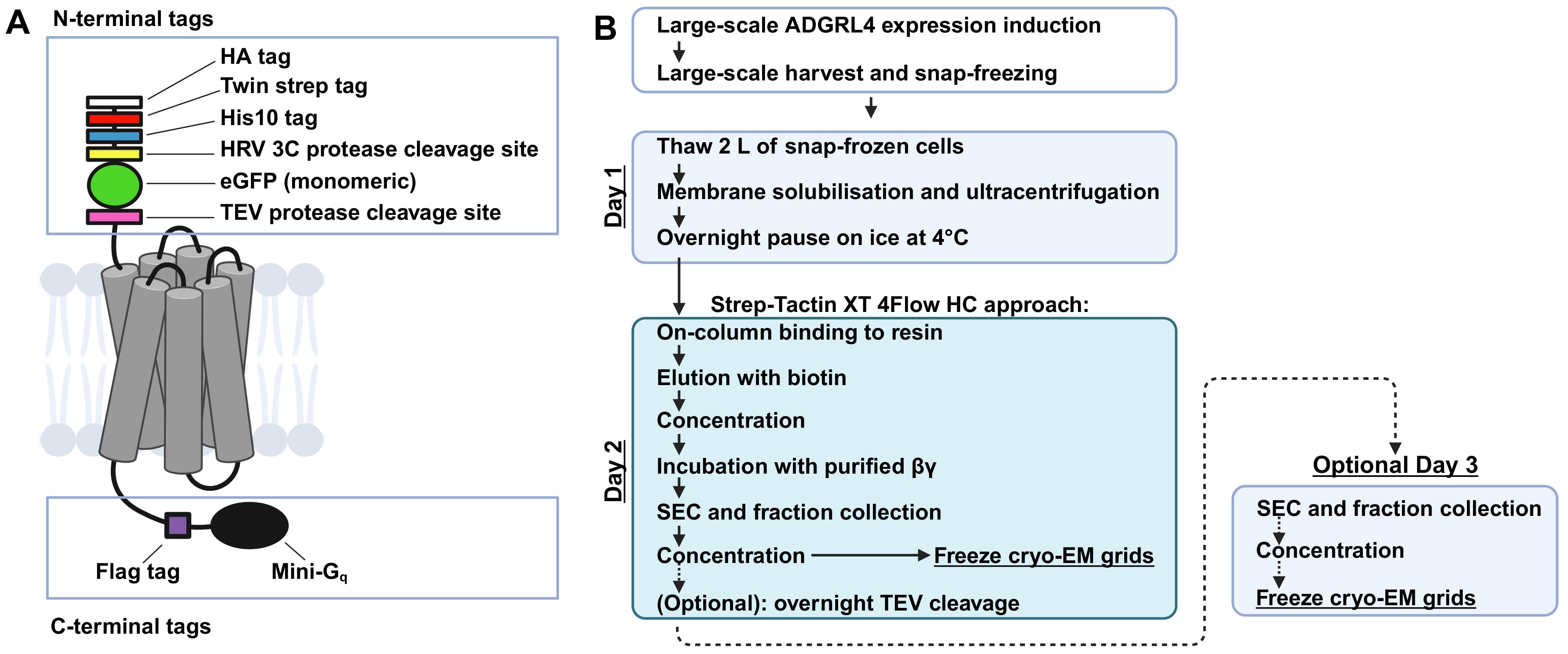
Graphical overview of ADGRL4 purification. (A) Schematic representation of active-state ADGRL4 illustrating the modular multifunctional tagging strategy. The N-terminal tag array provides orthogonal detection, purification, and protease cleavage options (HA, Twin-Strep, His10, HRV 3C cleavage site, monomeric eGFP, and TEV cleavage site). The C-terminus contains a FLAG tag followed by tethered mini-Gq. (B) Overview of the purification procedure. The steps shown for day 2 use Strep-Tactin XT 4Flow high-capacity resin, but any orthogonal purification approach compatible with the tagging strategy may be substituted. Day 3 (optional) outlines purification following overnight TEV cleavage to remove all N-terminal tags. This is optional, as the active-state ADGRL4 structure was determined using a protein that retained its N-terminal tags.
Background
Adhesion G protein-coupled receptors (aGPCRs) are a 32-member GPCR family in humans with diverse cellular roles [1].
ADGRL4 is an aGPCR implicated in tumour progression across multiple malignancies [2–22]. Using cryo-electron microscopy (cryo-EM), we recently determined the first high-resolution structure of active-state ADGRL4 to an overall nominal resolution of 3.1 Å [23], providing the first insights into its mechanism of activation. In the context of ADGRL4, the active state refers to a conformation of the receptor where the tethered agonist has bound to the orthosteric binding pocket, inducing a conformational change on the intracellular surface (outward movements of the intracellular ends of transmembrane helices 5 and 6) to form a cleft where the G protein Gq has coupled. (Subsequent use of the term active state in this protocol implies the above.) Active-state GPCRs are generally purified using detergent solubilisation in combination with engineered G proteins and/or stabilising binders. This has enabled numerous cryo-EM structures to be determined. Many different strategies have been developed, but explicit protocols for GPCR purification are generally unavailable, as they are usually terse descriptions in the methods section of a structural biology paper. An essential aspect of the purification procedure is the maintenance of the receptor complex during purification, which often requires careful choice of the GPCR construct expressed and the exact conditions [type and concentration of detergent(s), buffers, and salts] for the purification. The multi-parameter nature of purifying GPCRs thus makes it particularly challenging, particularly for researchers unfamiliar with the biochemistry of GPCRs; as such, we hope the detailed notes presented here will be useful. This protocol outlines the full workflow for purifying active-state ADGRL4 and can be completed in 2–3 days.
We developed our purification strategy for active-state ADGRL4 after first identifying its G protein coupling partner [23]. Using a NanoBiT split-luciferase complementation system, we found that ADGRL4 couples weakly to Gq. We subsequently observed that making an ADGRL4-mini-Gq chimera by genetically fusing mini-Gq to the C-terminus of active-state ADGRL4 markedly improved both receptor cell-surface expression and stability [23]. We therefore incorporated a C-terminal tethered mini-Gq (version R76 [24,25], capable of binding both βγ subunits of the heterotrimeric G protein complex; sequences in Dataset S1) to the constructs used for structural studies. Recruitment of βγ by the tethered mini-G increased the size of the receptor complex and introduced a distinct structural feature that served as a fiducial marker during cryo-EM particle alignment.
To maximise purification flexibility, we designed a modular system of N- and C-terminal detection, purification, and protease-cleavage tags, which did not adversely affect ADGRL4 cell-surface expression [23]. Our goal was to create a tagging architecture providing multiple orthogonal purification and detection options without requiring additional construct engineering. Most tags were positioned at the N-terminus immediately downstream of the native signal peptide sequence. The N-terminal cassette (Graphical overview; sequences in Dataset S1) consisted of (1) an HA tag for flow cytometry detection and HA-Trap purification, (2) a Twin-Strep tag for Strep-Tactin XT 4Flow high-capacity purification, (3) a His10 tag for nickel-nitrilotriacetic acid (Ni-NTA) purification, (4) an HRV 3c protease site to remove upstream tags, (5) monomeric eGFP (A206K) for flow cytometry detection and GFP-Trap purification, selected for its inability to self-dimerise [26], and (6) a TEV cleavage site to remove the entire N-terminal tag module. N-terminal tags were separated by GSG linkers. The TEV cleavage site was followed by a GSGGSG linker preceding the ADGRL4 sequence.
The C-terminus contained a FLAG tag for optional flow cytometry detection and FLAG-Trap purification, followed by the tethered mini-Gq (Graphical Overview; sequences in Dataset S1). To ensure adequate flexibility for the tethered mini-Gq, two GGSGSG linkers flanked the eight-residue FLAG tag. The C-terminal tethering of the mini-Gq was found to increase surface expression and improve stability of active-state ADGRL4 [23].
For structural studies, the ADGRL4 construct was packaged into lentivirus and used to generate an inducible HEK293 GnTI- TetR-based expression cell line, which was subsequently expanded at large scale [23]. The protocol for generating the ADGRL4 inducible HEK293 GnTI- TetR expression cell line is described in a companion Bio-protocol manuscript [27]. The use of a stable inducible cell line provides higher expression and better scalability compared with transient transfection and prevents the expression of intracellular, misfolded, and inactive receptor [28]. For purification, we selected the Twin-Strep tag in combination with Strep-Tactin XT 4Flow high-capacity resin, although any of the alternative orthogonal purification tags included in the design could be used interchangeably [23].
This protocol yields a high-purity active-state ADGRL4-mini-Gq-βγ complex suitable for cryo-EM grid preparation. Although optimised for ADGRL4, the tagging and mini-G protein tethering strategy and purification workflow are broadly applicable to other active-state aGPCRs and to weakly coupling or unstable GPCR-G protein complexes, particularly in cases where receptor instability has hindered structural characterisation.
Materials and reagents
Biological materials
1. Active-state ADGRL4 (version CTF2B) HEK293 GnTI- TetR stable cell line (stable suspension cell line with tetracycline inducible expression of the CTF2B active-state variant of ADGRL4. Available on request from Dr David Favara). Plasmids used to generate this stable inducible cell line (along with the detailed workflow for cell line development) are available on request from Dr David Favara.
2. Purified Gβγ dimer (69 mg/mL) [23] comprising human Gβ1 and Gγ2 (incorporating a C68S mutation to prevent post-translational lipidation). Expression and purification steps are detailed in the supplementary data section of [25]. Store at -80 °C. Available on request from Dr David Favara.
Reagents
1. Roche cOmplete, EDTA-free protease inhibitor cocktail tablets (Roche, catalog number: 11873580001); store at 4 °C
2. Phosphate-buffered saline (PBS), pH 7.4, 500 mL (Gibco, catalog number: 10010015)
3. Fetal bovine serum (FBS) tetracycline-free, 500 mL (Biosera, catalog number: FB-1001T/500); store at -20 °C
4. Trypan Blue solution, 0.4% (Gibco, catalog number: 15250061)
5. Anti-HA monoclonal antibody conjugated to APC, 200 μL (Miltenyi Biotec, catalog number: 130-123-553); store at 4 °C
6. HEPES buffer (molecular biology grade, ≥99%), 1 kg (Fisher BioReagents, catalog number: BP3101-1)
7. 4 N NaOH, 4 L (Supelco, catalog number: 1115845000)
8. 1 M NaOH, 500 mL (Supelco, catalog number: 79724-500ML)
9. PES membrane filter, 0.22 μm pore size (Millipore, catalog number: GPWP04700)
10. NaCl (Molecular biology grade, ≥99%), 500 g (Sigma-Aldrich, catalog number: S3014-500G)
11. MgCl2 hexahydrate (molecular biology grade, ≥99%), 500 g (Fisher BioReagents, catalog number: BP214500)
12. Glycerol (molecular biology grade, ≥99%), 1 L (Sigma-Aldrich, catalog number: G5516-1L)
13. Tris-(2-Carboxyethyl)phosphine, hydrochloride (TCEP) (molecular biology grade, ≥98%), 1 g (Sigma-Aldrich, catalog number: 75259-1G)
14. Apyrase (500 units/mL), 50 units (NEB, catalog number: M0398L); store at -20 °C
15. Phenylmethylsulfonyl fluoride (PMSF) protease inhibitor, 5 g (Thermo Scientific, catalog number: 36978)
16. Isopropanol (molecular biology grade, 99.5%), 1 L (Acros, catalog number: 10215331)
17. Lauryl maltose neopentyl glycol (LMNG), 25 g (Anatrace, catalog number: NG310 25 GM); store at -20 °C
18. Cholesteryl hemisuccinate tris salt (CHS), 5 g (Sigma-Aldrich, catalog number: C6013-5G); store at -20 °C
19. Benzonase nuclease, 25 KU (Millipore, catalog number: E1014-25KU); store at -20 °C
20. Standard SDS-PAGE reagents and pre-stained protein ladder (any suitable supplier)
21. Iba Lifesciences Strep-Tactin XT 4Flow high-capacity resin, 50 mL, 50% suspension (Iba Lifesciences, catalog number: 2-5030-025); store at 4 °C
22. Iba Lifesciences biotin, 5 g (Iba Lifesciences, catalog number: 2-1016-005); store at 4 °C
23. Iba Lifesciences buffer XT-R, 250 mL (Iba Lifesciences, catalog number: 2-1045-250); store at 4 °C
24. Purified TEV protease (4.4 mg/mL); expression and purification steps detailed in [29]. Alternatively, this can be purchased from NEB (NEB, catalog number: P8112S)
Solutions
1. Flow cytometry buffer (see Recipes)
2. 3 M NaCl (see Recipes)
3. 1 M MgCl2 (see Recipes)
4. 300 mM HEPES (see Recipes)
5. 50% glycerol (see Recipes)
6. 0.5 M TCEP (see Recipes)
7. 500 units/mL apyrase (see Recipes)
8. 0.5 M biotin (see Recipes)
9. 200 mM PMSF (see Recipes)
10. 5% LMNG (see Recipes)
11. 5% LMNG and 0.5% CHS (see Recipes)
12. Cell harvest buffer (see Recipes)
13. Solubilisation buffer (see Recipes)
14. Wash buffer (see Recipes)
15. Elution buffer (see Recipes)
16. SEC running buffer without glycerol (see Recipes)
17. SEC running buffer with 10% glycerol (see Recipes)
Recipes
1. Flow cytometry buffer
| Reagent | Final concentration | Quantity or volume |
|---|---|---|
| PBS | 98% | 19.6 mL |
| Tetracycline-free FBS | 2% | 0.4 mL |
| Total | n/a | 20 mL |
Note: Make up fresh and store at 4 °C. Keep on ice when in use.
2. 3 M NaCl
| Reagent | Final concentration | Quantity or volume |
|---|---|---|
| NaCl (≥99% pure) | 3 M | 175.32 g |
| Ultrapure water | see below | |
| Total | n/a | 1,000 mL |
a. Weigh 175.32 g of NaCl and dissolve in 700 mL of ultrapure water.
b. Bring to a total volume of 1,000 mL with ultrapure water.
c. Filter through a 0.22 μm pore size PES membrane filter into a sterile bottle and store at room temperature.
3. 1 M MgCl2
| Reagent | Final concentration | Quantity or volume |
|---|---|---|
| MgCl2 hexahydrate (≥99% pure) | 1 M | 20.33 g |
| Ultrapure water | see below | |
| Total | n/a | 100 mL |
a. Weigh 20.33 g of MgCl2 hexahydrate and dissolve in 50 mL of ultrapure water.
b. Bring to a total volume of 100 mL with ultrapure water. Store at room temperature.
4. 300 mM HEPES
| Reagent | Final concentration | Quantity or volume |
|---|---|---|
| HEPES (≥99% pure) | 300 mM | 71.5 g |
| Ultrapure water | see below | |
| NaOH used to titrate to pH 7.5 at 4 °C | see below | |
| Total | n/a | 1,000 mL |
a. Weigh 71.5 g of HEPES and dissolve in 900 mL of 4 °C ultrapure water. Keep on ice.
b. Measure pH and titrate carefully with 4 N NaOH (coarse titration) and then 1 M NaOH (fine titration) to pH 7.5 at 4 °C. Before titration, calibrate the pH meter with chilled buffers.
c. Bring to a total volume of 1,000 mL with ultrapure water.
d. Filter through a 0.22 μm pore size PES membrane filter into a sterile bottle and store at 4 °C.
5. 50% glycerol
| Reagent | Final concentration | Quantity or volume |
|---|---|---|
| Glycerol (≥99% pure) | 50% | 500 mL |
| Ultrapure water | 50% | 500 mL |
| Total | n/a | 1,000 mL |
a. Measure 500 mL of glycerol and mix with 500 mL of ultrapure water.
b. Autoclave at 121 °C for 15 min and store at 4 °C.
6. 0.5 M TCEP
| Reagent | Final concentration | Quantity or volume |
|---|---|---|
| TCEP (≥98% pure) | 0.5 M | 2.87 g |
| Ultrapure water | see below | |
| NaOH for titration to pH 7 at room temperature | see below | |
| Total | n/a | 20 mL |
a. Weigh 2.87 g of TCEP and dissolve it in 10 mL of ultrapure water.
b. Measure pH and titrate carefully with 4 N NaOH (coarse titration) and then 1 M NaOH (fine titration) to pH 7 at room temperature.
c. Bring to a total volume of 20 mL with ultrapure water.
d. Filter through a 0.22 μm pore size PES membrane filter into a sterile bottle.
e. Make up 1 mL aliquots and store at -20 °C.
Note: TCEP is a reducing agent that prevents the formation of non-native disulphide bonds. It is used to prevent deactivation of the TEV cysteine protease used for receptor cleavage.
7. 500 units/mL apyrase
Aliquoting for storage:
a. Source: apyrase 500 units/mL, 100 μL total, in 20 mM MES, 50 mM NaCl, 0.1 mM CaCl2, 1 mM DTT, 0.1% Tween-20, 50% glycerol.
b. Dispense 10 μL into ten 0.2 mL PCR tubes. Each aliquot contains 5 units apyrase at 500 units/mL.
c. Store at -20 °C.
Note: Apyrase degrades nucleoside triphosphates (ATP, GTP) and diphosphates (ADP, GDP) to their corresponding monophosphates and inorganic phosphate. This enzymatic activity depletes nucleoside tri- or diphosphates that could otherwise destabilise the active-state ADGRL4-mini-Gq-βγ complex. Activity typically requires Ca2+ or Mg2+. In this protocol, we use 1 mM MgCl2.
8. 0.5 M biotin
| Reagent | Final concentration | Quantity or volume |
|---|---|---|
| Biotin | 0.5 M | 5 g |
| Ultrapure water | see below | |
| NaOH and HCl to adjust pH | see below | |
| Total | n/a | 40.94 mL |
a. Place a glass beaker (≥50 mL) with a magnetic stir bar on a stir plate. Add 12 mL of ultrapure water and start stirring.
b. Weigh 5 g of biotin powder and add gradually while stirring to form a suspension. At this concentration, expect poor solubility below a pH of 7.5–8.
c. Measure pH and carefully titrate pH upward with 1 M NaOH in 0.5 mL increments. Switch to 100–200 μL increments of 1 M NaOH when the suspension becomes less thick and clearer. Allow time for each addition to fully mix before adding the next.
d. The suspension should become clear at pH 8–9.
e. Once suspension is completely clear, measure pH. The final target pH is between 8.2 and 8.4 If required, carefully titrate downward with 1 M HCl in 50–100 μL increments.
f. Once pH has settled at 8.2–8.4, bring to a final total volume of 40.94 mL with ultrapure water.
g. Filter through a 0.22 μm pore size PES membrane filter into a sterile bottle and store at 4 °C.
9. 200 mM PMSF
| Reagent | Final concentration | Quantity or volume |
|---|---|---|
| PMSF | 200 mM | 3.484 g |
| Isopropanol | to a 100 mL total volume | |
| Total | n/a | 100 mL |
a. In a fume hood, weigh 3.484 g of PMSF into a beaker.
b. Add 80 mL of isopropanol and mix by swirling to completely dissolve all PMSF.
c. Once fully dissolved, bring to a total volume of 100 mL with additional isopropanol.
d. Aliquot into 10 mL portions in 15 mL tubes and store at -20 °C, protected from light.
Caution: PMSF is toxic and corrosive and should be prepared in a fume hood. Ensure that it is always carefully handled with gloves.
Note: Only add PMSF to aqueous buffers immediately before use due it being unstable in water.
10. 5% LMNG
| Reagent | Final concentration | Quantity or volume |
|---|---|---|
| LMNG | 5% | 1 g |
| Ultrapure water | to a 20 mL total volume | |
| Total | n/a | 20 mL |
a. In a clear beaker or 50 mL tube, weigh 1 g of LMNG, mix with 10 mL of ultrapure water, and mix gently until dissolved. Avoid vigorous shaking to minimise bubble formation.
b. Bring to a final volume of 20 mL with additional ultrapure water.
c. Filter through a 0.22 μm pore size PES membrane filter and aliquot into 1.5 mL microcentrifuge tubes.
d. Store at -20 °C.
11. 5% LMNG and 0.5% CHS
| Reagent | Final concentration | Quantity or volume |
|---|---|---|
| LMNG | 5% | 2 g |
| CHS | 0.5% | 0.2 g |
| Ultrapure water | to a 40 mL total volume | |
| Total | n/a | 40 mL |
a. In a clear beaker or 50 mL tube, weigh 2 g of LMNG, mix with 20 mL of ultrapure water, and mix gently until dissolved. Avoid vigorous shaking to minimise bubble formation.
b. Weigh 0.2 g of CHS and add this to the LMNG solution.
c. Bring to a final volume of 40 mL with additional ultrapure water.
d. Gently mix overnight at 4 °C in a 50 mL tube using a tube roller.
e. Filter through a 0.22 μm pore size PES membrane filter into a sterile 50 mL tube.
f. Store at -20 °C.
12. Cell harvest buffer
| Reagent | Final concentration | Quantity or volume |
|---|---|---|
| 300 mM HEPES NaOH pH 7.5 | 10 mM | 3.33 mL |
| cOmplete EDTA-free protease inhibitor tablets (1 tablet for 50 mL) | 1× | 2 tablets |
| Ultrapure water | 1× | 96.67 mL |
| Total | n/a | 100 mL |
Note: Make up fresh on the day of harvesting and keep chilled at 4 °C. Dissolving cOmplete EDTA-free protease inhibitor tablets will require a stir bar for approximately 10 min at 4 °C.
13. Solubilisation buffer
| Reagent | Final concentration in 100 mL | Quantity or volume |
|---|---|---|
| 300 mM HEPES pH 7.5 | 20 mM | 6.67 mL |
| 3 M NaCl | 100 mM | 3.33 mL |
| 1 M MgCl2 | 10 mM | 1 mL |
| 50% glycerol | 20% | 40 mL |
| 0.5 M TCEP | 100 μM | 20 μL |
| 250 units/μL benzonase | 25 units/mL | 10 μL |
| 500 units/mL apyrase | 0.025 units/mL | 5 μL |
| cOmplete EDTA-free protease inhibitor tablets (1 tablet for 50 mL) | 1× | 2 tablets |
| 200 mM PMSF | 2 mM | 1 mL |
| Volume of cell pellet* | ~25 mL for 2 L of cells* | |
| Ultrapure water* | 3.965 mL | |
| Total | n/a | 80 mL |
* Include the volume of the thawed cell pellet in your calculation. When snap-freezing cells in liquid nitrogen, make a note of the volume of the pellet in the 50 mL centrifuge tube. For 2 L of HEK293 GnTI- TetR cells, this amounts to a ~25 mL volume.
Note: Make up fresh before use and keep on ice at 4 °C. Only add the benzonase and PMSF immediately before use. During solubilisation, 20 mL of 5% LMNG, 0.5% CHS is added to the above mixture, bringing the total volume to 100 mL (final detergent concentration being 1% LMNG, 0.1% CHS).
14. Wash buffer
| Reagent | Final concentration | Quantity or volume |
|---|---|---|
| 300 mM HEPES pH 7.5 | 20 mM | 6.67 mL |
| 3 M NaCl | 100 mM | 3.33 mL |
| 1 M MgCl2 | 10 mM | 1 mL |
| 50% glycerol | 20% | 40 mL |
| 0.5 M TCEP | 100 μM | 20 μL |
| 5% LMNG, 0.5% CHS | 0.02% LMNG, 0.002% CHS | 400 μL |
| cOmplete EDTA-free protease inhibitor tablets (1 tablet for 50 mL) | 1× | 2 tablets |
| 200 mM PMSF | 2 mM | 1 mL |
| Ultrapure water | 47.58 mL | |
| Total | n/a | 100 mL |
Note: Make up fresh before use and keep on ice at 4 °C.
15. Elution buffer
| Reagent | Final concentration | Quantity or volume |
|---|---|---|
| 300 mM HEPES pH 7.5 | 20 mM | 2.67 mL |
| 3 M NaCl | 100 mM | 1.33 mL |
| 1 M MgCl2 | 10 mM | 400 μL |
| 50% glycerol | 20% | 16 mL |
| 0.5 M TCEP | 100 μM | 8 μL |
| 5% LMNG, 0.5% CHS | 0.02% LMNG, 0.002% CHS | 160 μL |
| 0.5 M biotin | 50 mM | 4 mL |
| cOmplete EDTA-free protease inhibitor tablets (1 tablet for 50 mL) | 1× | 1 tablet |
| 200 mM PMSF | 2 mM | 400 μL |
| Ultrapure water | 15.032 mL | |
| Total | n/a | 40 mL |
Note: Make up fresh before use and keep on ice at 4 °C.
16. SEC running buffer without glycerol
| Reagent | Final concentration | Quantity or volume |
|---|---|---|
| 300 mM HEPES pH 7.5 | 20 mM | 13.33 mL |
| 3 M NaCl | 150 mM | 10 mL |
| 1 M MgCl2 | 10 mM | 2 mL |
| 0.5 M TCEP | 100 μM | 40 μL |
| 5% LMNG | 0.03% LMNG | 1.2 mL |
| Ultrapure water | 173.43 mL | |
| Total | n/a | 200 mL |
Note: Make up fresh before use and keep on ice at 4 °C.
17. SEC running buffer with 10% glycerol
| Reagent | Final concentration | Quantity or volume |
|---|---|---|
| 300 mM HEPES pH 7.5 | 20 mM | 3.33 mL |
| 3 M NaCl | 150 mM | 2.5 mL |
| 1 M MgCl2 | 10 mM | 500 μL |
| 50% glycerol | 10% | 10 mL |
| 0.5 M TCEP | 100 μM | 10 μL |
| 5% LMNG | 0.03% LMNG | 300 μL |
| Ultrapure water | 33.36 mL | |
| Total | n/a | 50 mL |
Note: Make up fresh before use and keep on ice at 4 °C.
Laboratory supplies
1. 96-well round (U) bottom plates (Thermo Scientific, catalog number: 163320)
2. 1 L polypropylene bottle assembly (Beckman Coulter, catalog number: C31597)
3. 50 mL centrifuge tubes (Corning, catalog number: CLS430828-100EA)
4. Ultracentrifuge tubes: Beckman Coulter PC tube with aluminium cap, 26.3 mL volume (Beckman Coulter, catalog number: 355618)
5. Econo-column chromatography column, 2.5 × 10 cm (Bio-Rad, catalog number: 7372512)
6. Microcentrifuge tubes, 1.5 mL (Pierce, catalog number: 69715)
7. Amicon Ultra-15 centrifugal filter 100 kDa MWCO (15 mL sample volume) (Millipore, catalog number: UFC910024)
8. Amicon Ultra-4 centrifugal filter 100 kDa MWCO (4 mL sample volume) (Millipore, catalog number: UFC810024)
9. Amicon Ultra-0.5 centrifugal filter 100 kDa MWCO (0.5 mL sample volume) (Millipore, catalog number: UFC510096)
10. Direct Detect assay-free cards, 50 cards (Merck, catalog number: DDAC00010)
11. UltrAuFoil R1.2/1.3 300 mesh Au grids (Quantifoil, catalog number: Q350AR13A)
Equipment
1. Flow cytometer (Sony, model: ID7000 Spectral Cell Analyzer)
2. Temperature-controlled floor-standing centrifuge for 1 L bottles (Beckman Coulter, Avanti JXN-26 Refrigerated Centrifuge, model: JXN-26)
3. Ultrapure water system (Sartorius, model: Arium Pro Ultrapure Water System)
4. Beckman Coulter Type 70 Ti fixed-angle rotor (Beckman Coulter, catalog number: 337922)
5. Temperature-controlled floor-standing ultracentrifuge (Beckman Coulter Optima L-100XP Ultracentrifuge, model: L-100XP)
6. End-over-end rotator (Elmi Rotamix RM1, catalog number: ROTAMIX RM1)
7. Shimadzu HPLC system (Shimadzu Corporation) comprising the following components: Shimadzu degasser module (DGU-20A), Shimadzu solvent delivery module (LC-20AD), Shimadzu autosampler module (SIL-20AC), Shimadzu column oven module (CTO-20AC), Shimadzu UV-detector module (SPD-20A), Shimadzu fluorescence detector module (RF-20A), and Shimadzu fraction collector module (FRC-40)
8. HPLC guard column, Agilent Bio SEC-5 guard column (300 Å, 7.8 × 50 mm, 5 μm) (Agilent, catalog number: 5190-2530)
9. HPLC column, Agilent Bio SEC-5 column (300 Å 4.6 × 300 mm, 5 μm) (Agilent, catalog number: 5190-2528)
10. Temperature-controlled benchtop 1.5 mL tube centrifuge (Eppendorf, model: 5418 R)
11. Protein detection system, Direct Detect infrared spectrometer (Merck, catalog number: DDHW00010)
12. Automated vitrification system for cryo-EM (FEI, model: Vitrobot Mark IV)
13. Ethane cryostat and temperature controller (MiTeGen, catalog number: ML-TCCS-001); design and operation are described in [30]
14. Glow-discharge plasma system (Fischione, catalog number: Model 1070)
Procedure
文章信息
稿件历史记录
提交日期: Dec 12, 2025
接收日期: Jan 26, 2026
在线发布日期: Feb 3, 2026
出版日期: Mar 5, 2026
版权信息
© 2026 The Author(s); This is an open access article under the CC BY-NC license (https://creativecommons.org/licenses/by-nc/4.0/).
如何引用
Favara, D. M. and Tate, C. G. (2026). Purification of the Active-State G Protein-Coupled Receptor ADGRL4 for Cryo-Electron Microscopy Using a Modular Tag System and a Tethered mini-Gq. Bio-protocol 16(5): e5617. DOI: 10.21769/BioProtoc.5617.
分类
生物化学 > 蛋白质 > 结构
生物物理学 > 电子冷冻断层扫描
您对这篇实验方法有问题吗?
在此处发布您的问题,我们将邀请本文作者来回答。同时,我们会将您的问题发布到Bio-protocol Exchange,以便寻求社区成员的帮助。