

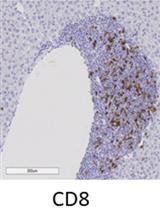
Immunohistochemical Staining of CD8α in Diabetic Mouse Kidney
糖尿病小鼠肾脏CD8α的免疫组化染色
发布: 2019年09月20日第9卷第18期 DOI: 10.21769/BioProtoc.3364 浏览次数: 3640
评审: Xiaoyi ZhengAnonymous reviewer(s)
参见作者原研究论文
The authors used this protocol in:
Sep 2018
Advertisement


Abstract
Immune cell infiltration, particularly cytotoxic CD8α lymphocyte infiltration, plays an important role in development of diabetic nephropathy. Although CD8α infiltration can be evaluated by its production of cytokines, its localization in the kidney is of particular importance. The current protocol describes CD8α immunostaining using a Vectastain ABC kit. This protocol works well with most commercially available antibodies, including CD8α antibodies in kidneys of diabetic mice.
Keywords: Diabetic nephropathy (糖尿病肾病)Background
Diabetic nephropathy (DN), a complication of diabetes, is the most common cause of end-stage renal disease. Pathological changes in DN are closely correlated with the degree of renal immune cell infiltration, particularly toxic CD8α lymphocyte infiltration. Therefore, a sensitive and reliable method to localize CD8α lymphocytes in the kidney will facilitate the research in diabetic nephropathy as well as other fields relating inflammation (Li et al., 2018).
Materials and Reagents
- Syringe
- Filter (Tisch Scientific, Millex, syringe-driven filter unit, 0.22 µm)
- Paraffin slides
- Glass staining jar (IMEB, SKU:SD-09)
- M.O.M Kit for detecting primary antibodies on mouse tissue (Vector Laboratories, Inc., catalog number: PK-2200)
Note: This Kit includes blocking solution and diluent for dilution of both primary and secondary antibodies (stored at 4 °C). - Biotinylated goat anti-mouse IgG antibody (Vector Laboratories, catalog number: BA-9200, stored at 4 °C)
- Vectastain Elite ABC HRP Kit (Vector Laboratories, catalog number: PK-6101, stored at 4 °C)
- Biotinylated goat anti-rat IgG antibody (Vector Laboratories, catalog number: BA-9400, stored at 4 °C)
- Rat anti-mouse CD8α antibody [AbD Serotec (now Bio-Rad Laboratories), catalog number: MCA2694, stored at -20 °C]
- Normal goat serum [Sigma-Aldrich (now Millipore, Sigma), catalog number: G9023-10ML, stored at -20 °C]
- Toluidine blue O (Sigma-Aldrich, catalog number: T3260-5G)
- 0.1 M Phosphate buffer (AlboChemicals, SKU: B0787MV4W2)
- Xylene
- Ethanol
- Methanol
- H2O2
- PBS
- Permount (Fisher Scientific, catalog number: SP15-100)
- Tris-HCl
- Blocking solution (see Recipes)
- Counterstain stock solution (see Recipes)
- Counterstain working solution (see Recipes)
- DAB solution (see Recipes)
Equipment
- Beakers (Nalgene, catalog number: 1542E06)
- Orbit shaker (Labline, catalog number: 3520)
- Regular light microscope (Wetzlar, Leitz Orthoplan)
- Oven
Procedure
文章信息
版权信息
© 2019 The Authors; exclusive licensee Bio-protocol LLC.
如何引用
Zhang, M. and Harris, R. C. (2019). Immunohistochemical Staining of CD8α in Diabetic Mouse Kidney. Bio-protocol 9(18): e3364. DOI: 10.21769/BioProtoc.3364.
分类
免疫学 > 免疫细胞染色 > 免疫检测
您对这篇实验方法有问题吗?
在此处发布您的问题,我们将邀请本文作者来回答。同时,我们会将您的问题发布到Bio-protocol Exchange,以便寻求社区成员的帮助。