现期刊物2026

卷册: 16, 期号: 6

Mar

20

Mar

5

Feb

20

Feb

5

Jan

20

Jan

5

生物信息学与计算生物学

利用 ECCFP 从 Nanopore 长读长测序数据中鉴定细胞外染色体环状 DNA 的生物信息学流程

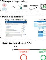
A Bioinformatics Workflow to Identify eccDNA Using ECCFP From Long-Read Nanopore Sequencing Data

利用 ECCFP 从 Nanopore 长读长测序数据中鉴定细胞外染色体环状 DNA 的生物信息学流程

WL Wang Li
BM Biyuan Miao
SW Shaogui Wan
79 Views
Mar 20, 2026
Extrachromosomal circular DNA (eccDNA) is a type of circular DNA that exists independently of chromosomes and has garnered significant attention in various fields, particularly in the context of smaller eccDNAs, which have considerable roles in gene regulation through various mechanisms. Current methods such as Circle-Seq and 3SEP can enrich small eccDNAs during sample preparation, but most bioinformatics pipelines remain challenging, exhibiting low accuracy and efficiency. This protocol describes the detailed workflow of a newly developed bioinformatics analysis pipeline, named EccDNA Caller based on Consecutive Full Pass (ECCFP), to accurately identify eccDNA from long-read Nanopore sequencing data. Compared to other pipelines, ECCFP significantly improves detection sensitivity, accuracy, and runtime efficiency. The process includes raw data quality control, trimming of adapters and barcodes, alignment to a reference genome, and identification of eccDNA, with detailed results encompassing accurate positioning of eccDNA, consensus sequences, and variants of individual eccDNA.

癌症生物学

Mag-Net 强阴离子交换技术用于分离卵巢癌腹水来源细胞外囊泡以开展蛋白质组学标志物发现

Mag-Net Strong Anion Exchange Enables Isolation of Ovarian Cancer Ascites Extracellular Vesicles for Proteomic Biomarker Discovery

Mag-Net 强阴离子交换技术用于分离卵巢癌腹水来源细胞外囊泡以开展蛋白质组学标志物发现

TC Tyler T. Cooper
96 Views
Mar 20, 2026
Extracellular vesicles (EVs) are nanoscale particles secreted by all cells and present in all biological fluids, where they carry molecular cargo reflective of health and disease states. Their diagnostic potential is often obscured by the high abundance of non-EV proteins and lipoproteins (e.g., albumin, apolipoproteins) that complicate proteomic analysis of primary biofluids, such as ascites fluid. Conventional isolation strategies face a persistent trade-off between EV purity and yield. To overcome this, a magnetic bead-based protocol (Mag-Net) to enrich EVs according to electrochemical surface charge using strong anion-exchange chemistry (SAX) was adapted for proteomics. Our workflow is specifically adapted to ascites fluid from human or murine sources. This approach effectively separates EVs from high-abundance proteins and lipoproteins, enabling proteomic profiling from as little as 2 μL of ascites fluid. Demonstrated in both murine and human ovarian cancer models, Mag-Net offers a reproducible, scalable, and automation-ready solution for EV isolation from various biofluids.
一种简单且低成本的三阴性乳腺癌细胞系(MDA-MB-231)球状体构建方法

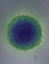
A Simple and Cost-Effective Method for Generating Spheroids From Triple-Negative Breast Cancer Cell Line (MDA-MB-231)

一种简单且低成本的三阴性乳腺癌细胞系(MDA-MB-231)球状体构建方法

Ramón Cervantes-Rivera Ramón Cervantes-Rivera
Luisa Nirvana González-Fernández Luisa Nirvana González-Fernández
AR Atalia Ziret Romero Rosas
SO Sandra Jetsamari Figueroa Ortíz
AO Alejandra Ochoa-Zarzosa
JL Joel E. López-Meza
57 Views
Mar 20, 2026
Breast cancer (BC) is the most frequently diagnosed malignancy in women and a leading cause of cancer-related mortality worldwide. Current clinical management relies on molecular classification—based on estrogen receptor (ER), progesterone receptor (PR), HER2, and Ki67 expression—to guide prognosis and therapy. Triple-negative breast cancer (TNBC), which lacks ER, PR, and HER2 expression, represents 15%–20% of cases and is characterized by aggressive behavior, early recurrence, and a paucity of targeted treatment options. These challenges underscore the urgent need for improved preclinical models that better recapitulate tumor biology to accelerate therapeutic discovery. While conventional monolayer (2D) cultures have contributed significantly to cancer research, they fail to mimic critical features of the three-dimensional (3D) tumor microenvironment (TME), thereby limiting clinical translation. To address this gap, 3D spheroid models have emerged as a powerful intermediary, more accurately replicating in vivo conditions such as cell–cell and cell–matrix interactions, nutrient and oxygen gradients, and the development of hypoxic cores. These features make spheroids a physiologically relevant platform for studying complex processes like metastasis, drug resistance, and treatment response. Here, we present a robust, simple, and cost-effective protocol for generating uniform 3D spheroids. Our method enables consistent monitoring of spheroid formation and growth over time, with quantitative, image-based size analysis to ensure reproducibility and scalability. Designed for flexibility, the protocol is broadly applicable across diverse cell types, effectively bridging the gap between traditional 2D cultures and complex in vivo studies. By providing an accessible and reliable model of the 3D TME, this protocol opens new avenues for high-throughput drug screening, mechanistic studies of tumor progression, and the advancement of personalized medicine strategies in breast cancer and beyond.

细胞生物学

基于径向分布分析的中心体蛋白定量方法

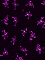
Radial Profile-Based Quantification of Centrosomal Proteins

基于径向分布分析的中心体蛋白定量方法

AW Alan Wainman
42 Views
Mar 20, 2026
Centrosomes are dynamic organelles critical for mitotic spindle assembly and cilia formation. Here, I describe a protocol for quantifying relative centrosomal protein abundance in Drosophila melanogaster embryos using radial profile analysis of fluorescence intensity. The method involves embryo collection, manual dechorionation, mounting for live imaging, confocal microscopy, and subsequent image analysis. Radial profiling allows quantification of relative protein abundance together with its spatial distribution at the centrosome, providing either relative or normalized intensity profiles. I then outline how this approach can be integrated with complementary techniques such as fluorescence recovery after photobleaching (FRAP) and super-resolution imaging, in this case, three-dimensional structured illumination microscopy (3D-SIM). Combining radial fluorescence profiling with these imaging modalities enables high-resolution, quantitative analysis of dynamic centrosome assembly in a genetically tractable system.

微生物学

一种用于单细胞水平定量寨卡病毒RNA的新型测序方法

A Novel Sequencing Method for Quantification of ZIKV RNA in Individual Cells

一种用于单细胞水平定量寨卡病毒RNA的新型测序方法

MH Min Hao
YW Yisong Wang
DD Dianyi Du
WY Wenrong Yang
QG Qiuzhe Guo
MT MingJing Tang
LL Libo Liu
WY Wei Yang
YL Yuxuan Liu
CL Chunyuan Luo
JC Jing Chen
PW Peigang Wang
JA Jing An
YZ Yang Zhou
40 Views
Mar 20, 2026
Single-cell RNA sequencing (scRNA-seq) is a powerful technique for exploring cellular heterogeneity and host–pathogen interactions. This protocol details the Zika virus (ZIKV)-targeted scRNA-seq workflow for preparing high-quality single-cell suspensions from the whole brain tissues of neonatal mice, high-quality single-cell sorting, cDNA reverse transcription, amplification, ZIKV enrichment and host transcriptome library preparation, and sequencing dataset integration in downstream analysis to complete the quantification of ZIKV RNA in individual cells.

分子生物学

基于胞嘧啶脱氨酶的基因组足迹分析方法(cFOOT-seq)用于检测转录因子占据情况

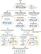
A Cytosine Deaminase–Based Genomic Footprinting Assay (cFOOT-seq) for Detecting Transcription Factor Occupancy

基于胞嘧啶脱氨酶的基因组足迹分析方法(cFOOT-seq)用于检测转录因子占据情况

MY Meng-Chen Yang
AW Ang Wu
HW Heng Wang
XL Xiaoyu Liu
JS Jiejun Shi
SG Shaorong Gao
JZ Jia-Min Zhang
84 Views
Mar 20, 2026
Transcription factors (TFs) regulate gene expression by binding to cis-regulatory elements in the genome. Understanding transcriptional regulation requires genome-wide characterization of TF occupancy across different chromatin contexts, yet simultaneous assessment of TF binding for multiple factors remains technically challenging. Here, we describe a detailed and reproducible protocol for cFOOT-seq, a cytosine deaminase–based genomic footprinting assay by sequencing, which enables antibody-independent, base-resolution profiling of chromatin accessibility, nucleosome organization, and TF occupancy. In cFOOT-seq, the double-stranded DNA (dsDNA) cytosine deaminase SsdAtox converts cytosine to uracil in accessible chromatin, whereas TF binding and nucleosome occupancy locally protect DNA from deamination. Using the FootTrack analysis framework, deamination patterns generated by cFOOT-seq are quantitatively analyzed to derive standardized footprint and chromatin organization profiles at base resolution across the genome. Because cFOOT-seq preserves genomic DNA integrity during deamination-based footprinting, it is compatible with ATAC-seq-based chromatin enrichment. ATAC-combined implementations reduce sequencing depth requirements and improve scalability for footprint-focused analyses, supporting applications in low-input and single-cell settings. This protocol provides a practical framework for genome-wide TF footprint profiling and can be readily applied to dissect gene regulatory mechanisms in development, immunity, and disease, including cancer.
RNA与蛋白质串联提取方法:最大化利用有限离体组织样本的实验平台

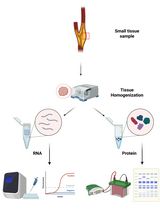
Tandem RNA and Protein Extraction: A Platform for Maximizing the Use of Limited Ex Vivo Tissue Samples

RNA与蛋白质串联提取方法:最大化利用有限离体组织样本的实验平台

CK Ciarán Kennedy
BM Braden Millar
LC Luke J. Conroy
MM Mariam Marai
MB Mary Barry
MO Martin O’Donohoe
OB Orina Belton
EB Eoin Brennan
CG Catherine Godson
Md Monica de Gaetano
46 Views
Mar 20, 2026
Human tissue samples represent the gold standard for obtaining clinically relevant and translatable insight into disease processes that in vitro systems cannot fully reproduce. However, patient-derived samples are often limited in size and availability, limiting the number of downstream assays that can be performed. To maximize the use of invaluable human samples, we present a protocol for the tandem extraction of high-quality RNA and protein from the same tissue section. This method has been optimized for 15–30 mg tissue sections, enabling more experimental conditions and technical replicates, while minimizing intrasample variability associated with heterogeneous tissues. This protocol also avoids potentially hazardous solvents present in phenol-chloroform-based methods such as TRIzol, providing a safer and more accessible workflow without compromising biomolecule integrity. This protocol was developed and validated using atherosclerotic plaque tissue from carotid endarterectomy, a very challenging tissue type to work with due to extensive calcification, necrosis, and limited surgical availability. We have also validated this method using mouse aortic tissue and cultured THP-1 cells, demonstrating its versatility across sample input types. As this protocol relies on standard column-based RNA extraction kits and commonly available reagents for protein precipitation and extraction, this methodology is widely accessible and easy to implement as a standard, streamlined workflow.

植物科学

拟南芥中纤维素合酶密度与运动速度测量的可重复性分析指南

A Guide to Reproducible Cellulose Synthase Density and Speed Measurements in Arabidopsis thaliana

拟南芥中纤维素合酶密度与运动速度测量的可重复性分析指南

JX Jan Y. Xue
SM Shawn D. Mansfield
AS A. Lacey Samuels
AS Arun Sampathkumar
RS René Schneider
139 Views
Mar 20, 2026
Cellulose synthase complexes (CSCs) play a central role in plant cell wall formation. Their dynamic behavior at the plasma membrane leads to the deposition of cellulose microfibrils into the apoplastic space, thereby shaping the architecture and mechanical properties of the cell wall. Although previous imaging studies have provided important insights into CSC dynamics and localization, standardized and reproducible workflows for quantitative measurements of CSC speed and density remain limited. Here, we present a reproducible live-cell imaging and analysis workflow for quantifying the speed and density of fluorescently labeled CSCs at the plasma membrane in Arabidopsis thaliana. The protocol integrates optimized spinning-disk confocal imaging, surface-based projection of z-stack recordings, automated detection of diffraction-limited CSCs foci, and kymograph-based speed measurements using freely available tools in Fiji. While selected steps, such as region of interest definition and parameter selection for spot detection or trajectory analysis, remain user-guided, these decisions are constrained to well-defined stages within an otherwise standardized pipeline, thereby reducing variability and improving reproducibility across experiments. The workflow has been validated across multiple tissues, reporter lines, genetic backgrounds, and perturbation conditions in Arabidopsis and enables robust comparative analysis of CSC dynamics. Beyond CSCs, this workflow is expected to be adaptable to other fluorescently labeled proteins that appear as diffraction-limited foci at or near the plasma membrane.
基于荧光的缺失等位基因特异性扩增(FAASA):用于高通量检测缺失等位基因的方法

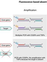
Fluorescence-Based Absent Allele-Specific Amplification (FAASA) for High-Throughput Detection of Absent Alleles

基于荧光的缺失等位基因特异性扩增(FAASA):用于高通量检测缺失等位基因的方法

KR Katherine L.D. Running
SS Sudeshi Seneviratne
ZZ Zengcui Zhang
GS Gurminder Singh
JF Jason D. Fiedler
JF Justin D. Faris
81 Views
Mar 20, 2026
In wheat and other crops, some genes display presence/absence variation, and it is occasionally beneficial to select for the absent allele to remove a functional gene. However, current high-throughput genotyping methods used to detect the absence of genes tend to be inconsistent and inconclusive. Kompetitive allele-specific PCR (KASP) and PCR allele competitive extension (PACE) are two well-established methods for allele-specific polymerase chain reaction (AS-PCR) assays, each using fluorescence resonance energy transfer (FRET) to generate a signal for each allele, typically targeting biallelic single-nucleotide polymorphisms. KASP and PACE methods are more difficult to apply to alleles with presence/absence variation because the lack of amplification of the absent allele is indistinguishable from a failed PCR. Here, we present a multiplex fluorescence-based absent allele–specific amplification (FAASA) method using the PACE marker system (compatible with KASP markers) to detect the absence of one particular or all alleles of a target sequence using a primer mix consisting of one target-specific primer pair (TSP) and a second primer set specific to a highly conserved endogenous gene known as a core gene–specific primer pair (CGSP). The forward primer of each pair is tagged with a 5′ terminal tail complementary to dye-labeled oligonucleotides in commercially available FRET cassettes. Lines that amplify only the core gene do not carry the target, while lines that amplify both the core gene and the target carry alleles of both the core gene and the target. The inclusion of the CGSPs allows researchers to confidently distinguish lines with absent alleles of the target from lines with failed PCR reactions, which can happen due to various reasons, including inadequate DNA quality or quantity.
利用RUBY可视化报告系统的快速大豆毛状根转化方法

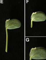
A Rapid and Visual Soybean Hairy Root Transformation Protocol Using the RUBY Reporter

利用RUBY可视化报告系统的快速大豆毛状根转化方法

ZZ Zhirui Zhang
QW Qiushi Wang
YG Yu Geng
JZ Jiuhai Zhao
59 Views
Mar 20, 2026
Agrobacterium rhizogenes–mediated hairy root transformation provides a rapid platform for gene function analysis prior to stable whole-plant transformation. However, most existing hairy root transformation methods rely on tissue culture and require chemical or fluorescence-based selection, which increases experimental complexity. Here, we describe a tissue culture–free soybean hairy root transformation protocol incorporating the RUBY visual reporter system. While this work does not introduce a new transformation concept, it presents a streamlined implementation of established soybean hairy root methodologies that emphasizes procedural simplicity, reduced handling, and faster access to functional root material. Transgenic roots expressing RUBY can be directly identified by red pigmentation with the naked eye. In RUBY-positive roots, candidate genes driven by the CaMV 35S promoter showed higher expression levels than those in empty-vector controls, indicating that the system supports effective gene expression. Using this procedure, clearly identifiable transgenic hairy roots can be obtained within 20 days. Overall, this protocol simplifies induction and screening while reducing operational complexity and equipment requirements.
利用规模化饲养飞虱在田间条件下实现 Fijivirus 的可控传播方法

Controlled Transmission of a Fijivirus Under Field Conditions Using Mass-Reared Planthoppers

利用规模化饲养飞虱在田间条件下实现 Fijivirus 的可控传播方法

AD Analía D. Dumón
MB Milene R. Barcenilla
JB Julieta B. Bariles
NP Nicolás A. Pereyra
SR Sandra M. Rodriguez
MM María F. Mattio
32 Views
Mar 20, 2026
Mal de Río Cuarto disease, caused by a Fijivirus, is a major constraint for maize production in Argentina. The traditional evaluation of resistant hybrids is limited by the low efficiency of natural virus transmission and the lack of standardized field inoculation methods. We developed a protocol that combines laboratory mass-rearing of the planthopper vector Delphacodes kuscheli with a controlled field transmission system. The method involves the synchronized production of large insect populations, acquisition of viruliferous vectors under controlled conditions, and their safe transport to the field using specialized containers. Transmission is achieved through individual cages placed on maize seedlings, ensuring high inoculation pressure under field-like conditions. This protocol enables reliable and reproducible virus transmission, facilitating large-scale screening of maize hybrids and other cereals. Its main advantages are the high throughput of vector production, improved transmission efficiency, and adaptability to diverse experimental designs.